. 2025 Jun 7;26(12):5487.
doi: 10.3390/ijms26125487.

Sponge bHLH Gene Expression in Xenopus laevis Disrupts Inner Ear and Lateral Line Neurosensory Development and Otic Afferent Pathfinding

Affiliations

Sponge bHLH Gene Expression in Xenopus laevis Disrupts Inner Ear and Lateral Line Neurosensory Development and Otic Afferent Pathfinding

Karen L Elliott et al. Int J Mol Sci. .

Abstract

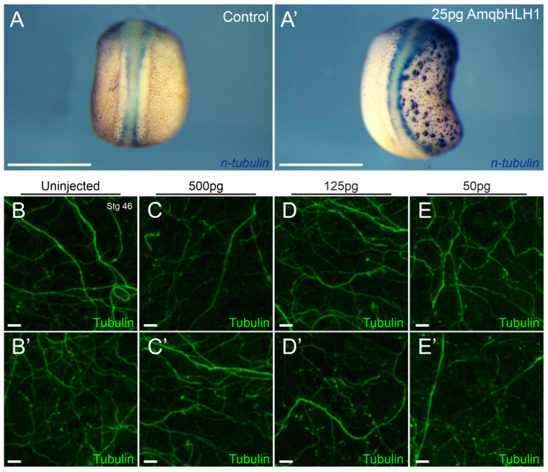
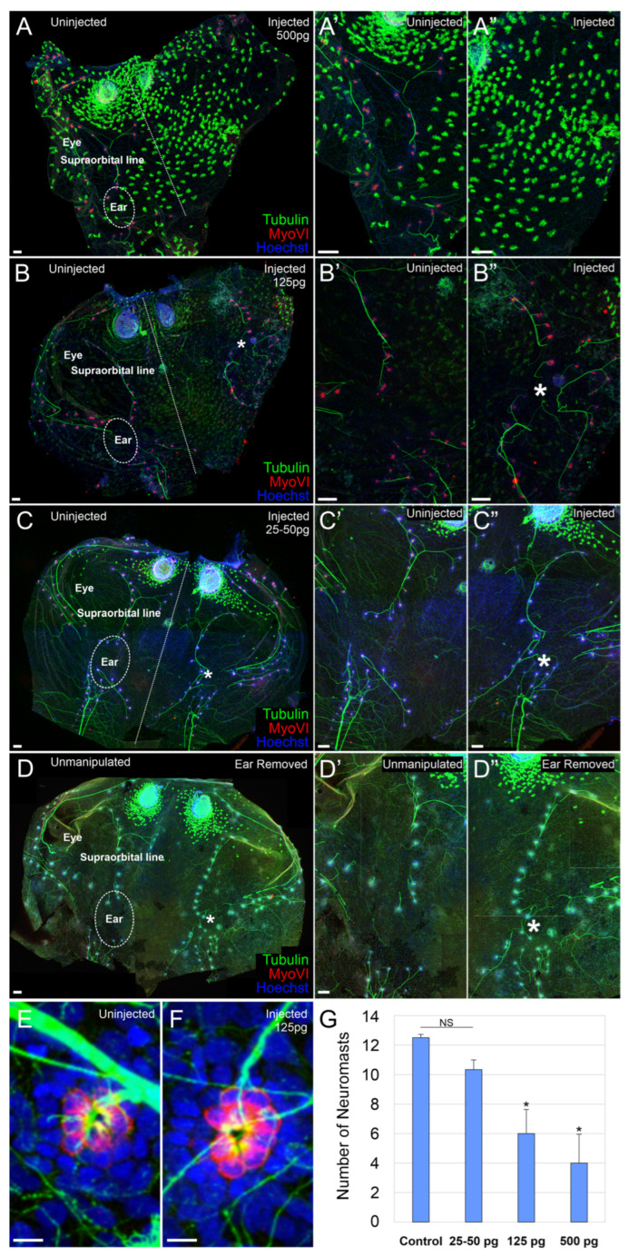
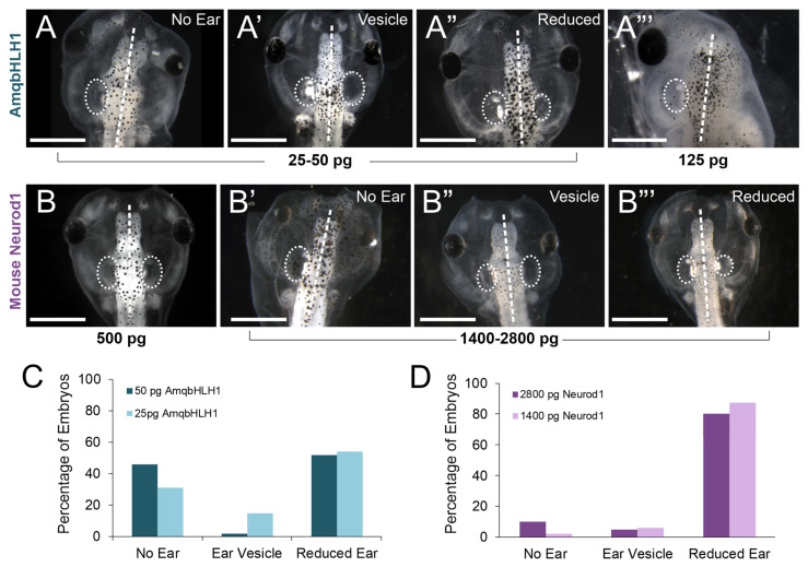
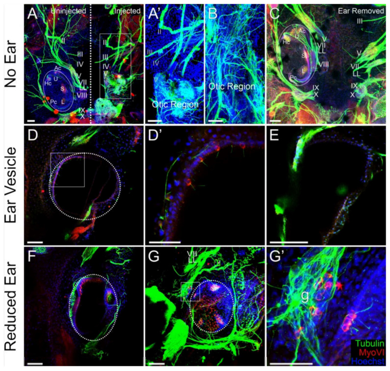
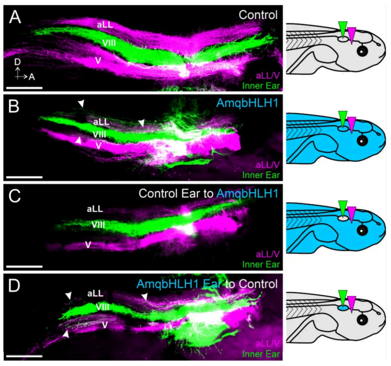
Basic helix-loop-helix (bHLH) transcription factors, such as those in the atonal family, are important in cellular fate determination. The expression of the sponge ortholog of the atonal bHLH gene family, AmqbHLH1, in Xenopus laevis previously resulted in the formation of ectodermal ectopic neurons. However, the extent to which these neurons persist through development and the effects on the inner ear and lateral line, which require a critical level and timing of bHLH genes, remains unexplored. To test these long-term effects, we injected various concentrations of AmqbHLH1 mRNA into X. laevis embryos and assessed neurosensory development at developmental stages coinciding with fully developed neurosensory structures. The expression of AmqbHLH1 mRNA in X. laevis resulted in a dose-dependent reduction in or loss of ears and the lateral line system without eliminating ectopic neurons. At the lowest concentrations examined, we found that inner ear neurosensory development consisted sometimes of only a few scattered hair cells in a single-layer epithelium. Furthermore, low concentrations of AmqbHLH1 mRNA affected inner ear afferent guidance. Our data suggest that the AmqbHLH1 gene has some anti-neurosensory abilities in frogs and that the overexpression of a single gene may not be sufficient for stable long-term transdifferentiation in cells.

Keywords: Neurod1; atonal; bHLH; inner ear; lateral line; neurosensory development.


Conflict of interest statement

The authors declare no conflict of interest.

Figures

Figure 1
Figure 1
AmqbHLH1 mRNA does not have a long-term ectopic neuron effect in the ectoderm. (A,A’) In situ hybridization for n-tubulin expression in control neurula-stage embryos (A) and in embryos injected on the right side with 25 pg AmqbHLH1 mRNA (A’) (eight of eleven animals showed positive staining at 25 pg of stage 20); (BE’) 100 µm × 100 µm areas of representative cranial ectoderm of stage 46 tadpoles stained with acetylated tubulin antibody. Images show non-lateral line neurons present in the skin in uninjected halves (B,B’) and in halves injected with 500 pg AmqbHLH1 mRNA (C,C’), 125 pg AmqbHLH1 mRNA (D,D’), and 50 pg AmqbHLH1 mRNA (E,E’). Scale bar is 1 mm in (A,A’) and 10 μm in (BE’).
Figure 2
Figure 2
AmqbHLH1 mRNA affects the lateral line. (AC) Immunohistochemistry using antibodies against acetylated tubulin (green) and MyoVI (red) from an animal in which the right half was injected with 500 pg (A), 125 pg (B), or 25–50 pg (C) AmqbHLH1 mRNA. (A’,A”) Higher magnification of the uninjected and injected halves, respectively, of (A), showing the absence of a supraorbital lateral line on the injected side. (B’,B”). Higher magnification of the uninjected and injected halves, respectively, of (B), showing some abnormal development in the lateral line. (C’,C”). Higher magnification of the uninjected and injected halves, respectively, of (C), showing near-normal development in the lateral line. (D) Immunohistochemistry using antibodies against acetylated tubulin (green) and myoVI (red) from an animal in which the right ear was physically removed at stage 25–27. (D’,D”) Higher magnification of the unmanipulated and ear-removed halves, respectively, of (D). For both types of manipulations in (C,D), there are no lateral line neuromasts over the ear (circled); however, when the ear was absent/reduced following AmqbHLH1 mRNA injection or absent following physical ear removal, there was an expansion of lateral line neuromasts into the region previously occupied by the ear (white asterisks). (E) Single neuromast from an uninjected side. (F) Single neuromast from an animal injected with 125 pg AmqbHLH1 mRNA. (G) Number of neuromasts in the supraorbital line of the lateral line system, ± SEM, * p < 0.05; NS, not significant. Scale bars are 100 μm in (AD”) and 10 μm in (E,F). Skins were counterstained with the nucleus marker Hoechst, dash line in (B,C) show the boundary of left and right side. (E) at any of the doses of AmqbHLH1 mRNA (hair cells were counted in six neuromasts from each of the four animals).
Figure 3
Figure 3
Phenotype assessment of ear development following AmqbHLH1 or mouse Neurod1 mRNA expression. (AA”) Stage 46 Xenopus laevis tadpoles showing the no-ear, ear-vesicle, and reduced-ear phenotypes, respectively, following the injection of either 25 pg or 50 pg of AmqbHLH1 mRNA into the right half of the animal. (A’”) Stage 46 X. laevis tadpole showing no ear, as well as no eye, on the AmqbHLH1 mRNA-injected (right) side. (B) Stage 46 X. laevis tadpole showing no effect following injection of mouse Neurod1 mRNA. (B’B’”) Stage 46 X. laevis tadpoles showing the no-ear, ear-vesicle, and reduced-ear phenotypes, respectively, following the injection of either 1400 pg or 2800 pg of mouse Neurod1 mRNA into the right half of the animal. Scale bars are 1 mm. (C) Percentage of embryos with the no-ear, ear-vesicle, or reduced-ear phenotypes for 25 pg and 50 pg of AmqbHLH1 mRNA. (D) Percentage of embryos with the no-ear, ear-vesicle, or reduced-ear phenotypes for 1400 pg and 2800 pg of mouse Neurod1 mRNA. Dot circles indicate the size of ears, dash line indicate the left/right side. Scale bars in (AB’’’) are 1 mm.
Figure 4
Figure 4
Low doses of AmqbHLH1 mRNA disrupt the neurosensory development of the inner ear. (AG’) Immunohistochemistry using antibodies against acetylated tubulin (green) and MyoVI (red) in stage 46 tadpoles unilaterally injected with 25 pg or 50 pg of AmqbHLH1 mRNA. (A,B) Immunohistochemistry confirming the complete loss of the inner ear and instead showing a mass of neurons in the otic region on the injected (right) side in two animals in which no ear was detected. The anterior is at the top in all images. (A’) Higher magnification of the boxed area in (A). (C) An animal in which an ear was physically removed reveals an approximation of pre- and post-otic cranial ganglia but no overall disorganization. (D,E) Immunohistochemistry revealed limited (D) to no (E) neurosensory development in two animals with an ear vesicle on the injected side. (D’) Higher magnification of the boxed area in (D), showing a few scattered hair cells in a single-layer epithelium. (F,G) Immunohistochemistry showed a lack of segregation of the utricle–saccule–lagena in two animals with reduced ears. (G’) Higher magnification of the boxed area in (G), showing the presence of myoVI-positive cells in the otic ganglia (g). Abbreviations: Ac, anterior canal; Hc, horizontal canal; L, lagena; Pc, posterior canal; S, saccule; U, utricle; II, optic nerve; III, oculomotor nerve; IV, trochlear nerve; V, trigeminal nerve; VII, facial; LL, lateral line; VIII, vestibular nerve; IX, glossopharyngeal nerve; X, vagus nerve. Dot circles indicate the size of ears, dash line indicate the left/right side, box is larger image (D,D’,G,G’). Scale bars are 100 μm in (A,A’,BD,EG) and 50 μm in (D’,G’).
Figure 5
Figure 5
AmqbHLH1 mRNA disrupts the central pathfinding of inner ear afferents. (A) Lipophilic dyes implanted into the ear (green) and into the trigeminal and anterior lateral line nerves (magenta) of a control animal demonstrate normal central projections. Grey and light blue background indicates tadpoles with or without mRNA. The inner ear afferent projections to the vestibular nucleus (VIII) are bordered dorsally by anterior lateral line afferents (aLLs) and ventrally by trigeminal afferents (V). The dorsal area is at the top and the anterior is on the right in all images. (B) Lipophilic dye implantation in the ear of an animal injected with AmqbHLH1 mRNA reveals inner ear projections that not only project to the vestibular nucleus but also to the lateral line and trigeminal nuclei (arrowheads). (C) Lipophilic dye implantation in the ear from a control animal transplanted into an animal injected with AmqbHLH1 mRNA reveals normal central projections. (D) Lipophilic dye implantation in the ear of an animal injected with AmqbHLH1 mRNA transplanted into a control animal reveals inner ear projections that not only project to the vestibular nucleus but also to the lateral line and trigeminal nuclei (arrowheads). Scale bars are 100 μm. Diagrams represent treatment (gray, control; blue, AmqbHLH mRNA-injected), and colored wedges represent lipophilic dye placement (green, inner ear; magenta, anterior lateral line and trigeminal nerves).

Similar articles

References

    1. Imayoshi I., Kageyama R. bHLH Factors in Self-Renewal, Multipotency, and Fate Choice of Neural Progenitor Cells. Neuron. 2014;82:9–23. doi: 10.1016/j.neuron.2014.03.018. - DOI - PubMed
    1. Kageyama R., Shimojo H., Ohtsuka T. Dynamic control of neural stem cells by bHLH factors. Neurosci. Res. 2018;138:12–18. doi: 10.1016/j.neures.2018.09.005. - DOI - PubMed
    1. Elliott K.L., Pavlínková G., Chizhikov V.V., Yamoah E.N., Fritzsch B. Development in the Mammalian Auditory System Depends on Transcription Factors. Int. J. Mol. Sci. 2021;22:4189. doi: 10.3390/ijms22084189. - DOI - PMC - PubMed
    1. Zine A., Fritzsch B. Early Steps towards Hearing: Placodes and Sensory Development. Int. J. Mol. Sci. 2023;24:6994. doi: 10.3390/ijms24086994. - DOI - PMC - PubMed
    1. Butts J.C., Wu S.R., Durham M.A., Dhindsa R.S., Revelli J.P., Ljungberg M.C., Saulnier O., McLaren M.E., Taylor M.D., Zoghbi H.Y. A single-cell transcriptomic map of the developing Atoh1 lineage identifies neural fate decisions and neuronal diversity in the hindbrain. Dev. Cell. 2024;59:2171–2188.e2177. doi: 10.1016/j.devcel.2024.07.007. - DOI - PubMed

MeSH terms

Substances

LinkOut - more resources