. 2025 Jun 8;26(12):5504.
doi: 10.3390/ijms26125504.

Mitochondrial Transfer from Human Platelets to Rat Dental Pulp-Derived Fibroblasts in the 2D In Vitro System: Additional Implication in PRP Therapy

Affiliations

Mitochondrial Transfer from Human Platelets to Rat Dental Pulp-Derived Fibroblasts in the 2D In Vitro System: Additional Implication in PRP Therapy

Koji Nishiyama et al. Int J Mol Sci. .

Abstract

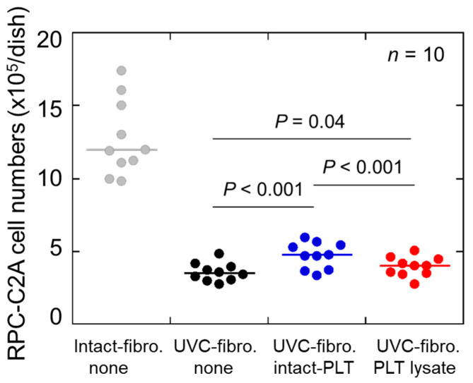
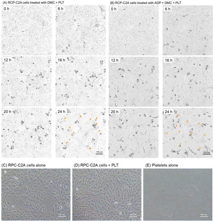
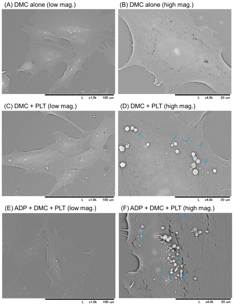
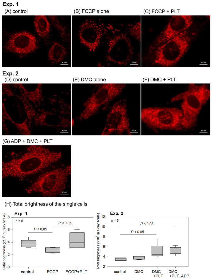
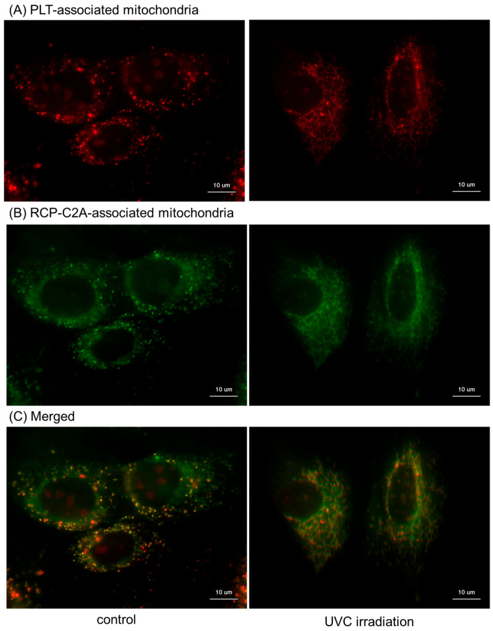
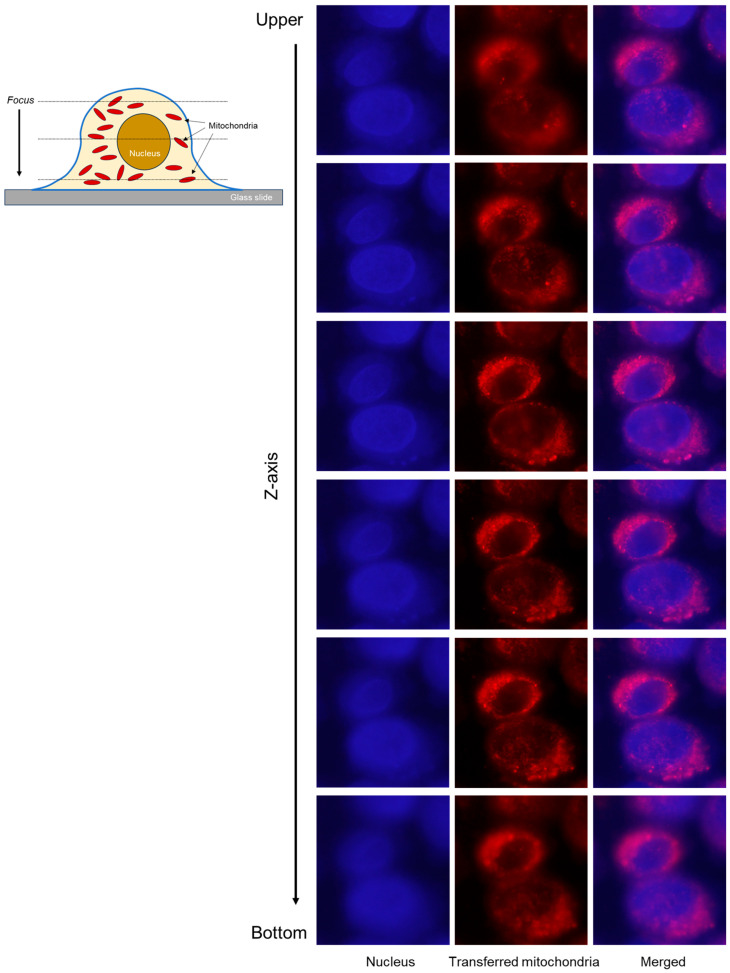
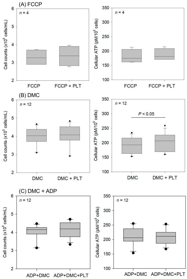
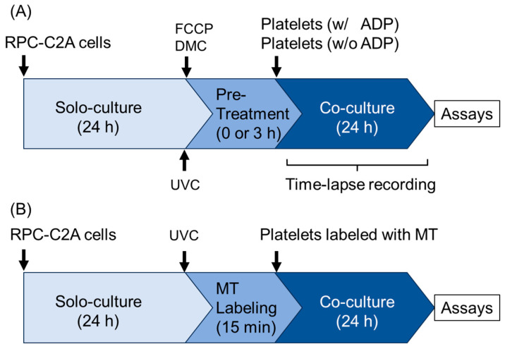
Platelet mitochondria have recently been increasingly considered "co-principal" along with platelet growth factors to facilitate tissue regeneration in platelet-rich plasma therapy cooperatively. To develop a convenient method to test this potential, we examined mitochondrial transfer using a simple two-dimensional culture system. Living human platelets were prepared from PRP obtained from 12 non-smoking healthy male adults (age: 28-63 years) and suspended in medium. Platelet lysates were prepared from sonicated platelet suspensions in PBS. After treatment with ultraviolet-C irradiation, a mitochondrial respiration inhibitor, or a synchronized culture reagent, rat dental pulp-derived fibroblasts (RPC-C2A) were co-cultured with platelets or platelet lysates for 24 h. Mitochondrial transfer was evaluated by visualization using a fluorescent dye for mitochondria or an antibody against human mitochondria. Ultraviolet-C-irradiated cells substantially lost their viability, and treatment with living platelets, but not platelet lysates, significantly rescued the damaged fibroblasts. Fibroblast mitochondria appeared to increase after co-culture with resting platelets. Although more microparticles existed around the platelets on the fibroblast surface, the activated platelets did not show significant increases in any parameters of mitochondrial transfer. This simple co-culture system demonstrated mitochondrial transfer between xenogeneic cells, and this phenomenon should be considered as an additional implication in PRP therapy.

Keywords: fibroblasts; in vitro; mitochondria; platelet-rich plasma; platelets; transfer.


Conflict of interest statement

The authors declare no conflicts of interest.

Figures

Figure 1
Figure 1
Rescuing effects of living platelets (PLTs) on UVC-irradiated RPC-C2A cells. After 24 h solo-culture and medium exchange, RPC-C2A cells were irradiated with UVC and cultured with platelets for 24 h. At the end of the treatment, cells were gently washed and counted for analysis of cell viability. Cell numbers without UVC irradiation were inserted in a blue square with a blue error bar. n = 10.
Figure 2
Figure 2
Time-course changes in platelet counts (observed as tiny black dots in extracellular spaces) in co-culture with RPC-C2A cells (orange arrows indicate some samples). After pretreatment with demecolcine (DMC), the cells were treated with living platelets in the absence (A) or presence (B) of ADP. Similar results were obtained from three independent experiments. Orange arrows indicate representative RCP-C2A cells at various growth phases. (C) RPC-C2A cells alone at 20 h. (D) RPC-C2A cells + platelets at 20 h. (E) Platelets alone at 20 h. Scale bar = 100 μm.
Figure 3
Figure 3
Photomicrographs by scanning electron microscopy of platelets and their microparticles on the surfaces of RPC-C2A cells. After pretreatment with demecolcine, cells were treated with living platelets (PLTs) for 24 h in the absence (C,D) or presence (E,F) of ADP. Control cells were pretreated with demecolcine (DMC) in the control (A,B). Similar findings were obtained from the other three independent experiments. Based on the difference in number, light blue arrows indicate representative microvesicles, probably released from activated platelets. Scale bar = 100 μm (low magnification) or 20 μm (high magnification).
Figure 4
Figure 4
Effects of living platelets (PLTs) on the number of intact mitochondria indicated by membrane potential staining in RPC-C2A cells. In Experiment 1, after pretreatment with FCCP, cells were treated with living PLTs for 24 h in the absence (B,C) of ADP. In control (A), cells were not treated with inhibitors or PLTs. In Experiment 2, after pretreatment with demecolcine (DMC), cells were treated with living PLTs for 24 h in the absence (E,F) or presence of ADP (G). In control (D), cells were not treated with PLTs. Scale bar = 10 μm. (H) Quantification of the total brightness of each representative single cell. n = 5.
Figure 5
Figure 5
Effects of living platelets (PLTs) on the distribution of human platelet mitochondria in RPC-C2A cells. After pretreatment with demecolcine (DMC), the cells were treated with living PLTs for 24 h in the absence (B) or presence (C,D) of ADP. In the control (A), cells were pretreated with demecolcine alone. Human mitochondria and fibroblast nuclei were stained with anti-human mitochondria antibody, followed by visualization with secondary antibody (red), and DAPI (dark blue), respectively. Similar findings were obtained from the other three independent experiments. Scale bar = 10 μm.
Figure 6
Figure 6
Distribution of platelet mitochondria transferred to RPC-C2A cells. Platelet and fibroblast mitochondria were labeled with MitoTracker Orange CMTMRos ((A): red) and MitoTracker Green FM ((B): green), respectively. After 24 h co-cultures, cells were gently washed, fixed with 10% neutralized formaldehyde, and examined. To observe individual distribution, both images were merged (C). Scale bar = 10 μm.
Figure 7
Figure 7
Z-stack images of RPC-C2A cells co-cultured with platelets, in which mitochondria were labeled with MitoTracker Orange CMTMRos (red). After 24 h of co-culture without other treatments, fibroblasts were enzymatically detached, pasted on glass slides using cytospin, fixed, and stained with DAPI (dark blue). As illustrated in the upper left panel, serial images were obtained using a fluorescence microscope along the Z-axis with an appropriate step size (approximately 0.15–0.2 μm) without changing the X- and Y-axes. Both images were merged to observe the mitochondrial distribution. Scale bar = 10 μm.
Figure 8
Figure 8
Effects of living platelets (PLTs) on intact RPC-C2A cell counts and cellular ATP levels. After pretreatment with FCCP (A) or demecolcine (DMC) (B,C), the cells were treated with living PLTs for 24 h in the absence (A,B) or presence (C) of ADP. n = 4 (A) and 12 (B,C).
Figure 9
Figure 9
Experimental protocols for cell treatments. (A) Protocols for cell survival assay, time-lapse recording, mitochondrial membrane potential staining, immunofluorescence staining, and SEM examination. (B) Protocol for MitoTracker labeling assay. FCCP: carbonyl cyanide 4-(trifluoromethoxy)phenylhydrazone, DMC: demecolcine, UVC: ultraviolet-C, MT: MitoTracker. “Solo-culture” represents the culture of RPC-C2A cells alone without platelets.

Similar articles

References

    1. Kawase T. Platelet-rich plasma and its derivatives as promising bioactive materials for regenerative medicine: Basic principles and concepts underlying recent advances. Odontology. 2015;103:126–135. doi: 10.1007/s10266-015-0209-2. - DOI - PubMed
    1. Kawase T. A Strategic and Worldwide Cooperative Challenge Required for the Next Generation of Platelet Concentrates. Int. J. Mol. Sci. 2022;23:3437. doi: 10.3390/ijms23073437. - DOI - PMC - PubMed
    1. Kawase T., Mubarak S., Mourão C.F. The Platelet Concentrates Therapy: From the Biased Past to the Anticipated Future. Bioengineering. 2020;7:82. doi: 10.3390/bioengineering7030082. - DOI - PMC - PubMed
    1. Knightly N., Lee C., O’Brien L., Qayyum T., Hurley C., Kelly J. Role for platelet rich plasma as an adjuvant therapy in wound healing and burns. Eur. J. Plast. Surg. 2023;46:465–474. doi: 10.1007/s00238-023-02050-8. - DOI
    1. El-Sharkawy H., Kantarci A., Deady J., Hasturk H., Liu H., Alshahat M., Van Dyke T.E. Platelet-rich plasma: Growth factors and pro- and anti-inflammatory properties. J. Periodontol. 2007;78:661–669. doi: 10.1902/jop.2007.060302. - DOI - PubMed

LinkOut - more resources