Review
. 2025 Jun 9;26(12):5508.
doi: 10.3390/ijms26125508.

Unveiling GABA and Serotonin Interactions During Neurodevelopment to Re-Open Adult Critical Periods for Neuropsychiatric Disorders

Affiliations
Review

Unveiling GABA and Serotonin Interactions During Neurodevelopment to Re-Open Adult Critical Periods for Neuropsychiatric Disorders

Emanuela Beretta et al. Int J Mol Sci. .

Abstract

The mature brain is the result of a complex neurodevelopmental process resulting from interweaved mechanisms and involving early genetic and microenvironmental factors shaped by patterns of spontaneous electrical activity. During postnatal development, the immature brain undergoes experience-dependent structural and functional shaping and modifications during critical period (CP) time windows to achieve the full maturation of brain functions. Plasticity is higher during neurodevelopmental CP windows and is limited in the adult brain, including during neuropsychiatric disorders. Notably, the neurotransmitters γ-aminobutyric acid (GABA) and serotonin are two fundamental players controlling and modulating, respectively, brain plasticity in the developing and adult brain. Therefore, acquiring insights into the roles played by GABA and serotonin in regulating CP plasticity might hold potential for pharmacologically re-opening CP windows in adult life, with the aim of providing therapeutic intervention for neurological and neuropsychiatric disorders.

Keywords: GABA; adult plasticity; cognition; neurodevelopment; neuropsychiatric disorders; plasticity; serotonin.


Conflict of interest statement

The authors declare no conflicts of interest.

Figures

Figure 2
Figure 2
Development of the serotonergic system. (a) Schematic cartoons of rodent sagittal brain sections at embryonic days 13 (E13, (top)) and 17 (E17, (bottom)), showing the emergence and clustering of serotonergic neurons. By E17, serotonergic neurons are grouped into caudal (B1–B3, light green) and rostral (B4–B9, purple) clusters. Serotonergic fibers destined for the forebrain (purple arrow) travel along the median forebrain bundle (MFB) and originate from the dorsal and median raphe. Serotonergic fibers destined to the spinal cord (light-green arrow) arise from the B1–B3 clusters (red circles) and travel along the medial longitudinal fasciculus (MLF) [adapted from [60]. (b) Serotonin innervation in the central nervous system. (Top): schematic diagram showing the major rostral (purple) and caudal (green) serotonergic projections. (Bottom): serotonergic innervation of the spinal cord, with the raphe pallidus (B1) projecting to the ventral horn, the raphe obscurus (B2) innervating the intermediate zone, and the raphe magnus (B3) projecting to the dorsal horn of the spinal cord. Yellow circles indicate motor neurons [adapted with permission under the terms of the Creative Commons Attribution License, available at https://creativecommons.org/licenses/by/4.0/legalcode.en (accessed on 27 May 2025) (CC-BY License) from [60]. Created with BioRender.com.
Figure 3
Figure 3
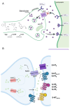
The serotonergic system. (A) Schematic cartoon of a serotonergic synapse with pre- and postsynaptic sites. The tryptophan (Tryp) introduced to the diet is hydroxylated into 5-hydroxytryptophan (5-HTP) by tryptophan 5-hydroxylase 2 (THP 2), and then decarboxylated into 5-hydroxytryptamine (5-HT) or serotonin by the aromatic L-amino acid decarboxylase enzyme (DDC). Once packed into the vesicles, serotonin is released into the synaptic cleft and binds postsynaptic receptors (i.e., 5-HT1A, 5-HT1B, 5-HT1D, and 5-HT2A,C). Serotonin is transported into the presynaptic terminal thanks to the action of the serotonin reuptake transporter (SERT) present in the plasma membrane. Here, 5-HT is broken down by MAO (monoamine oxidase) and, for future release, is repackaged into vesicles by isoform 2 of the vesicular monoamine transporter (VMAT2). Created with BioRender.com. (B) Schematic cartoon of the 5-HT receptor families. MAO: metabolic enzymes monoamine oxidase; SERT: serotonin reuptake transporter; VMAT2: vesicular monoamine transporter 2. [adapted with permission under the terms of the Creative Commons Attribution License, available at https://creativecommons.org/licenses/by/4.0/legalcode.en (accessed on 27 May 2025) (CC-BY License) from [78].
Figure 4
Figure 4
Schematic representation of radial and tangential migration. (Left): There are two primary types of neuronal migration: radial and tangential. In radial migration, pyramidal (glutamatergic) neurons travel perpendicularly to the ventricular surface along radial glial fibers (yellow). In contrast, during tangential migration, inhibitory (GABAergic) neurons follow paths that are parallel to the ventricular surface and perpendicular to the radial glial fibers (purple) [adapted with permission under the terms of the Creative Commons Attribution License, available at https://creativecommons.org/licenses/by/4.0/legalcode.en (accessed on 27 May 2025) (CC-BY License) from [101]. (Right): Specifically, excitatory pyramidal neurons are generated from glial progenitor cells (RGPs) in the VZ and migrate radially toward the cortical plate (CP), following the apical RGP process. MZ: marginal zone; CP: cortical plate; IZ: intermediate zone; SVZ: subventricular zone. Non-pyramidal inhibitory neurons, on the other hand, arise from the medial ganglionic eminence (MGE), migrate tangentially in streams, and later invade the cortex. Created with BioRender.com.
Figure 1
Figure 1
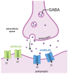
Synaptic and extra-synaptic GABAergic transmission. In the presynaptic terminal, the neurotransmitter GABA is packed within synaptic vesicles. When GABA is released into the synaptic cleft, it activates synaptic fast-phasic GABAA receptors (sGABAARs, in blue). In the extra-synaptic zone, low ambient GABA concentration binds slow extra-synaptic GABAA receptors (eGABAARs, in light green). Extra-synaptic GABAA receptors desensitize less rapidly than synaptic GABAA receptors, eliciting a constant tonic current in the neurons. Created with BioRender.com.

Similar articles

References

    1. Bystron I., Blakemore C., Rakic P. Development of the human cerebral cortex: Boulder Committee revisited. Nat. Rev. Neurosci. 2008;9:110–122. doi: 10.1038/nrn2252. - DOI - PubMed
    1. Marín O., Rubenstein J.L.R. Cell Migration in the Forebrain. Annu. Rev. Neurosci. 2003;26:441–483. doi: 10.1146/annurev.neuro.26.041002.131058. - DOI - PubMed
    1. Homem C.C.F., Repic M., Knoblich J.A. Proliferation control in neural stem and progenitor cells. Nat. Rev. Neurosci. 2015;16:647–659. doi: 10.1038/nrn4021. - DOI - PMC - PubMed
    1. Deidda G., Biazzo M. Gut and Brain: Investigating Physiological and Pathological Interactions Between Microbiota and Brain to Gain New Therapeutic Avenues for Brain Diseases. Front. Neurosci. 2021;15:753915. doi: 10.3389/fnins.2021.753915. - DOI - PMC - PubMed
    1. Murphy T.H., Corbett D. Plasticity during stroke recovery: From synapse to behaviour. Nat. Rev. Neurosci. 2009;10:861–872. doi: 10.1038/nrn2735. - DOI - PubMed

LinkOut - more resources