. 2025 Jun 9;26(12):5520.
doi: 10.3390/ijms26125520.

Cells of the Maternal-Fetal Interface May Contribute to Epidural-Related Maternal Fever After Administration of Ropivacaine: The Role of Phosphatases DUSP9 and PHLPP1

Affiliations

Cells of the Maternal-Fetal Interface May Contribute to Epidural-Related Maternal Fever After Administration of Ropivacaine: The Role of Phosphatases DUSP9 and PHLPP1

Florian Horn et al. Int J Mol Sci. .

Abstract

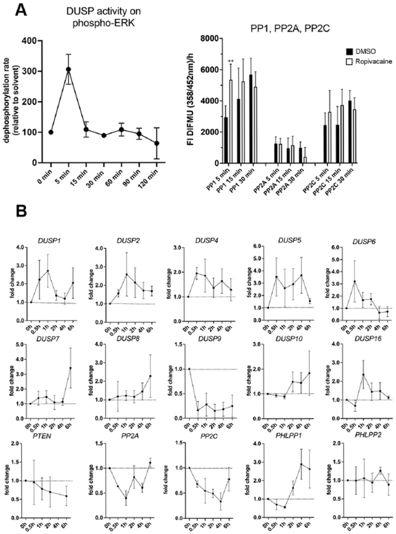
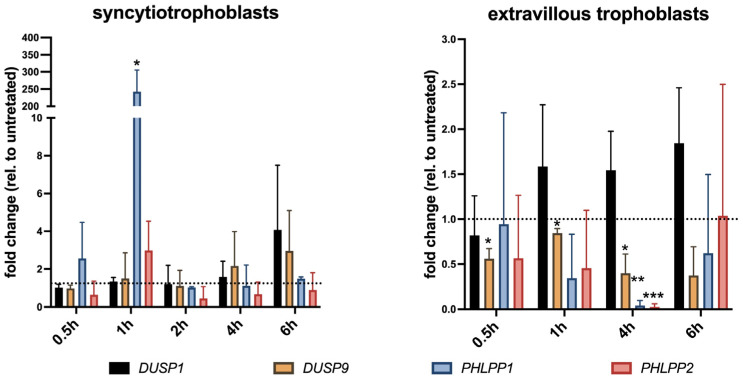
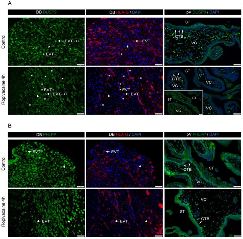
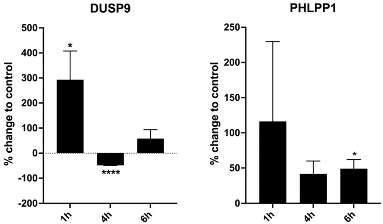
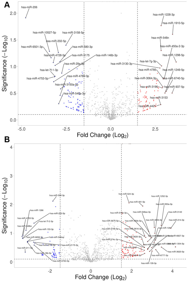
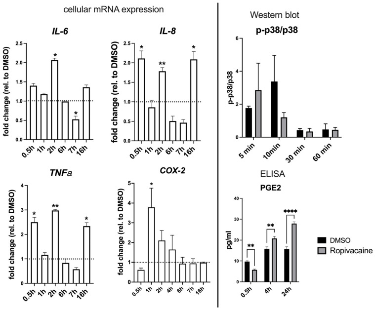
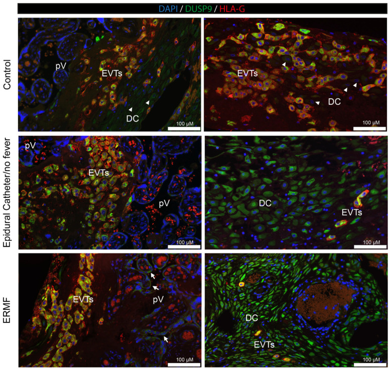
Epidural-related maternal fever (ERMF) occurs with significant incidence in women receiving local anesthetics such as ropivacaine via epidural catheter for pain relief during labor. The causal mechanism behind this phenomenon is still not fully resolved, but evidence suggests that these anesthetics cause sterile inflammation. In this observational study, we investigated a possible contributory role of the dual-specificity phosphatase-9 (DUSP9) controlling the activity of mitogen-activated protein kinases (MAPK), and also PH-domain and Leucine-rich repeat phosphatase (PHLPP) regulating AKT kinases. The data show that ropivacaine differentially affects the expression of these phosphatases in distinct cell types of the umbilical cord and placenta. The gene expression of DUSP9 was almost completely switched off in the presence of ropivacaine in HUVECs and extravillous trophoblasts for up to 6 h, while the expression of PHLPP1 was upregulated in HUVECs and syncytiotrophoblasts. Extravillous trophoblasts were identified as a source of pro-inflammatory mediators and regulatory miRNAs in response to ropivacaine. Placentae at term exhibited a distinct DUSP9 expression pattern, whether the patients belonged to the control group or received epidural analgesia with or without elevated body temperature. The observed data imply that ropivacaine induces complex effects on the MAPK and AKT pathways at the feto-maternal interface, which contribute to the ERMF phenomenon.

Keywords: DUSP9; ERMF; PHLPP1; dual-specificity phosphatase; epidural analgesia; epidural-related maternal fever.


Conflict of interest statement

The authors declare no conflicts of interest.

Figures

Figure 1
Figure 1
Effects of ropivacaine on different phosphatase activities and expression of MAPK-DUSPs and AKT-regulating phosphatases in HUVECs. (A) Phosphatase activity assays of MAPK-DUSPs with ERK as the substrate and protein phosphatases-1, -2A, and -2C in HUVECs upon exposure to 0.01% (100 μg/mL) ropivacaine. (B) qPCR analyses of gene expression of MAPK-DUSPs (MKP) and phosphatases regulating the AKT pathway after exposure of HUVECs to 0.01% ropivacaine. The plots show mean values ± S.D. from n = 3 different preparations of HUVECs. 2 Asterisks (**) means p smaller than 0.01.
Figure 2
Figure 2
qPCR gene expression analysis for DUSP1, DUSP9, PHLPP1, and PHLPP2 in cultured syncytiotrophoblasts and extravillous trophoblasts upon exposure to 0.01% ropivacaine. DMSO-stimulated cells served as vehicle controls. Cytotrophoblasts were allowed to fuse for 72 h before stimulation. Non-stimulated controls were arbitrarily set to 1 (= no change; dotted line). Bars indicate mean values ± SD of two isolations (6th to 9th week of pregnancy, n = 4 placentae per isolation), measured in duplicate. * p < 0.05; ** p < 0.01; *** p < 0.001 (Wilcoxon test).
Figure 3
Figure 3
Immunofluorescence analyses showing representative examples of DUSP9 and pan-PHLPP expression in first-trimester decidual and placental explant cultures in the absence (control) or presence of 0.01% ropivacaine. Explants exposed to DMSO served as the vehicle control. (Left panel) Representative pictures of DUSP9 (A) and pan-PHLPP (B) staining (green) of decidua basalis (9th week of pregnancy; n = 2 preparations). Corresponding serial sections stained with HLA-G (red) to visualize EVTs and DAPI (blue; nuclei) are depicted in the middle panel. Arrows indicate representative DUSP9- or PHLPP-positive EVTs (HLA-G-positive). Arrowheads mark examples of DUSP9- or PHLPP-positive decidual cells lacking HLA-G expression. Asterisks show representative EVTs with low DUSP9 expression (HLA-G-positive). (Right panel) Staining of first-trimester placental villi (9th week of pregnancy; n = 2 preparations) using DUSP9 or PHLPP (A,B, respectively) antibodies, counterstained with DAPI (blue; nuclei). Arrows indicate examples of cytotrophoblasts. Bars = 50 µm. CTB, cytotrophoblast; DB, decidua basalis; EVTs, extravillous trophoblasts; pV, placental villus; ST, syncytiotrophoblast; VC, villous core.
Figure 4
Figure 4
Western blot analysis of first-trimester explant tissue of total decidua basalis exposed to 0.01% ropivacaine for 1 h, 4 h, and 6 h developed with antibodies for DUSP9 and PHLPP1. Tissue samples of similar weight were exposed to 0.01% ropivacaine and DMSO as solvent in parallel. Equal amounts of protein were processed for Western blot analysis. Bars indicate percentage change in ropivacaine-exposed tissue relative to vehicle-treated tissue as mean ± SD. Data are from n = 3 replicates. * p < 0.05; **** p < 0.0001 (Wilcoxon Test).
Figure 5
Figure 5
Volcano plots showing altered expressions of miRNAs in extravillous trophoblasts upon ropivacaine exposure. Cells were exposed to 0.01% ropivacaine in the cell culture for (A) 0.5 h and (B) 4 h. miRNAs were quantified using small RNA sequencing and expressed as log2fold change relative to the solvent (DMSO) for the same time span.
Figure 6
Figure 6
Inflammatory responses in cultured extravillous trophoblasts upon exposure to 0.01% ropivacaine. Due to higher sensitivity, the expression of TNFα, IL-6, IL-8, and COX-2 was analyzed by qPCR on the mRNA level. The p-p38/p38 ratio was determined from the intensities of the respective bands in Western blots, and PGE2 levels were analyzed in the supernatant by ELISA. Black bars represent treatment with DMSO, grey bars represent treatment with 0.01% ropivacaine in DMSO. All data were determined from n = 3 replicates and are shown as mean ± SD. * p < 0.05; ** p < 0.01; **** p < 0.0001 (two-way ANOVA, multiple comparisons).
Figure 7
Figure 7
Immunofluorescence analysis of DUSP9 in term placental tissues from women in the absence (control) or presence of an epidural catheter/ropivacaine administration, with or without fever (ERMF and epidural catheter/no fever, respectively). n = 3 per group. Two representative pictures of different parts of the placenta are shown. The left column displays decidual tissue in close proximity to the placental villi, and the right column shows deeper invasion sites. Sections were stained with DUSP9 (green), HLA-G (red) to visualize EVTs, and DAPI (blue; nuclei). Double-positive EVTs appear orange. Arrows indicated DUSP9-positive syncytiotrophoblasts of placental villi. Arrowheads point towards examples of DUSP9-negative decidual cells. Bars = 100 μm. DC, decidual cells; EVTs, extravillous trophoblasts; pV, placental villi.

Similar articles

References

    1. Beilin Y., Halpern S. Focused review: Ropivacaine versus bupivacaine for epidural labor analgesia. Anesth. Analg. 2010;111:482–487. doi: 10.1213/ANE.0b013e3181e3a08e. - DOI - PubMed
    1. Fusi L., Steer P.J., Maresh M.J., Beard R.W. Maternal pyrexia associated with the use of epidural analgesia in labour. Lancet. 1989;1:1250–1252. doi: 10.1016/S0140-6736(89)92341-6. - DOI - PubMed
    1. Patel S., Ciechanowicz S., Blumenfeld Y.J., Sultan P. Epidural-related maternal fever: Incidence, pathophysiology, outcomes, and management. Am. J. Obstet. Gynecol. 2023;228:S1283–S1304.e1. doi: 10.1016/j.ajog.2022.06.026. - DOI - PubMed
    1. Yancey M.K., Zhang J., Schwarz J., Dietrich C.S., 3rd, Klebanoff M. Labor epidural analgesia and intrapartum maternal hyperthermia. Pt 1Obstet. Gynecol. 2001;98:763–770. doi: 10.1097/00006250-200111000-00010. - DOI - PubMed
    1. Wohlrab P., Boehme S., Kaun C., Wojta J., Spittler A., Saleh L., Knofler M., Markstaller K., Klein K.U., Tretter V. Ropivacaine Activates Multiple Proapoptotic and Inflammatory Signaling Pathways That Might Subsume to Trigger Epidural-Related Maternal Fever. Anesth. Analg. 2020;130:321–331. doi: 10.1213/ANE.0000000000004402. - DOI - PubMed

MeSH terms

LinkOut - more resources