Review
. 2025 Jun 10;26(12):5539.
doi: 10.3390/ijms26125539.

Precision Oncology Framework Using Circulating Tumor Cells

Affiliations
Review

Precision Oncology Framework Using Circulating Tumor Cells

Fumihiko Kakizaki et al. Int J Mol Sci. .

Abstract

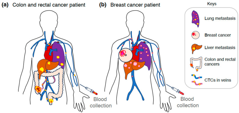
Circulating tumor cells (CTCs) are multifaceted biomarkers with significant potential for precision oncology, offering opportunities to refine diagnoses and personalize treatments across various cancer types, including colorectal and breast cancer. CTC assays serve as reliable prognostic indicators, even during chemotherapy and/or molecularly targeted therapies. Notably, CTCs exhibit heterogeneity that gradually develops during carcinogenesis and becomes more pronounced in advanced disease stages. These intra- and intertumoral heterogeneities pose challenges, particularly when drug-resistant clones emerge following therapy. The dynamic behavior of CTCs provides valuable insights into treatment response and prognosis. Extensive efforts have led to the development of technologies for effective CTC isolation, accelerating their clinical implementation. While both CTC and circulating tumor DNA (ctDNA) tests offer prognostic value, they reflect different aspects of tumor biology: CTC counts indicate tumor progression, while ctDNA levels correlate with tumor burden. The combined analysis is expected to yield complementary insights. CTC tests are feasible in general hospitals and may serve as tumor markers comparable to, or even superior to, conventional markers such as carcinoembryonic antigen (CEA) and carbohydrate antigen 19-9 (CA19-9) for colorectal cancer, and CA15-3 for breast cancer. Early incorporation of CTC tests into routine blood panels appears to be a rational and promising approach.

Keywords: CTC; breast cancer; colorectal cancer; ctDNA; precision oncology.


Conflict of interest statement

The authors declare the following conflicts of interest: F.K., H.M. (Hisatsugu Maekawa), H.M. (Hiroyuki Miyoshi), Y.T., and K.O. (Kazutaka Obama) are affiliated with an endowed chair established through funding received from AFI Corporation, Kyo Diagnostics K.K., and SCREEN Holdings Co., Ltd. F.K., H.M. (Hisatsugu Maekawa), H.M. (Hiroyuki Miyoshi), Y.T., and K.O. (Kazutaka Obama) received research funds from AFI Corporation, Kyo Diagnostics K.K., and SCREEN Holdings Co., Ltd. K.O. (Kyoichi Oshiro) is an employee of AFI Corporation.

Figures

Figure 2
Figure 2
Heterogeneity in CTC transcriptomes of metastatic colorectal and breast cancers. (a,b) Heatmaps showing CTC transcriptomic profiles in metastatic colorectal (n = 59; panel (a)) and breast cancers (n = 339; panel (b)) [25,49,50,51,52,53,54,64]. In panel (b), ctcRbase integrates seven datasets (GSE111065, GSE51827, GSE55807, GSE67939, GSE75367, GSE86978, and GSE41245) as previously described [64]. Panels (a) and (b) present expression values in transcripts per million (TPM) and fragments per kilobase of transcript per million mapped reads (FPKM), respectively. These heatmaps were reconstructed by the authors based on data reported in previous studies [25,49,50,51,52,53,54,64]. As only limited genomic data on CTCs was available in these datasets, gene mutation data is not presented.
Figure 1
Figure 1
Overview of tumor dissemination and blood collection for CTC analysis. Colorectal (yellow, panel (a)) and breast (pink, panel (b)) carcinomas invade surrounding tissues, reach nearby veins (red purple, or blue), and enter the bloodstream as CTCs (yellow or pink circles). In panel (a), CTCs of colorectal cancer are frequently transported to the liver (brown) via the mesenteric and portal veins (red-purple) and subsequently reach the lungs (dark purple). In contrast, lower rectal cancer cells primarily metastasize to the lungs (dark purple) via the inferior rectal vein (blue), inferior vena cava, and heart. In panel (b), breast cancer cells are transported to the bones (not shown), lungs (dark purple), and liver (brown) through the venous circulation and heart.

References

    1. Le Tourneau C., Borcoman E., Kamal M. Molecular profiling in precision medicine oncology. Nat. Med. 2019;25:711–712. doi: 10.1038/s41591-019-0442-2. - DOI - PubMed
    1. Yates L.R., Seoane J., Le Tourneau C., Siu L.L., Marais R., Michiels S., Soria J.C., Campbell P., Normanno N., Scarpa A., et al. The European Society for Medical Oncology (ESMO) Precision Medicine Glossary. Ann. Oncol. 2018;29:30–35. doi: 10.1093/annonc/mdx707. - DOI - PubMed
    1. Mateo J., Steuten L., Aftimos P., André F., Davies M., Garralda E., Geissler J., Husereau D., Martinez-Lopez I., Normanno N., et al. Delivering precision oncology to patients with cancer. Nat. Med. 2022;28:658–665. doi: 10.1038/s41591-022-01717-2. - DOI - PubMed
    1. Veyrune L., Naumann D.N., Christou N. Circulating Tumour Cells as Prognostic Biomarkers in Colorectal Cancer: A Systematic Review. Int. J. Mol. Sci. 2021;22:3437. doi: 10.3390/ijms22083437. - DOI - PMC - PubMed
    1. Hu M., Wang Z., Wu Z., Ding P., Pei R., Wang Q., Xing C. Circulating tumor cells in colorectal cancer in the era of precision medicine. J. Mol. Med. 2022;100:197–213. doi: 10.1007/s00109-021-02162-3. - DOI - PMC - PubMed

LinkOut - more resources