. 2025 Jun 10;26(12):5540.
doi: 10.3390/ijms26125540.

SARS-Cov-2 Replication in a Blood-Brain Barrier Model Established with Human Brain Microvascular Endothelial Cells Induces Permeability and Disables ACE2-Dependent Regulation of Bradykinin B1 Receptor

Affiliations

SARS-Cov-2 Replication in a Blood-Brain Barrier Model Established with Human Brain Microvascular Endothelial Cells Induces Permeability and Disables ACE2-Dependent Regulation of Bradykinin B1 Receptor

Sharton Vinicius Antunes Coelho et al. Int J Mol Sci. .

Abstract

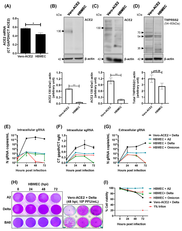
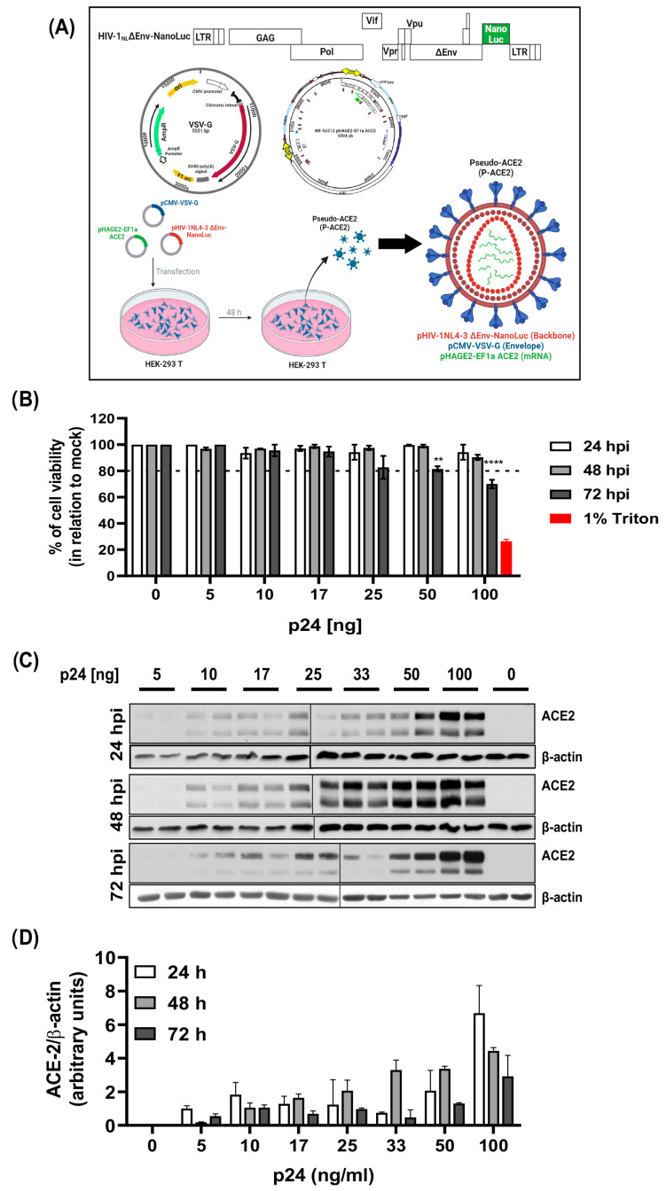
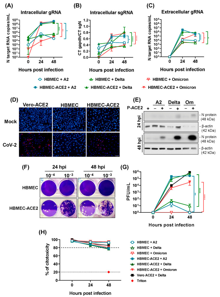
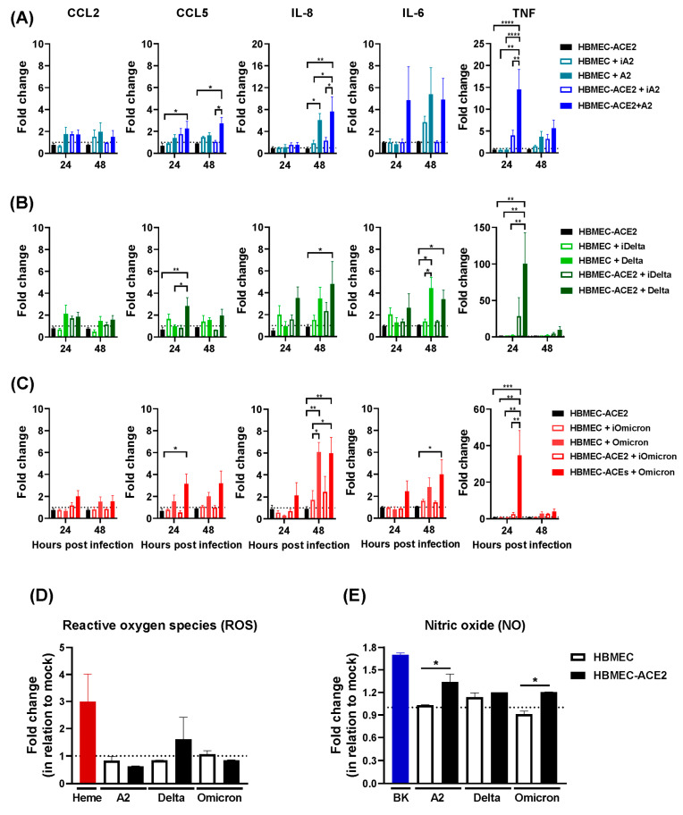
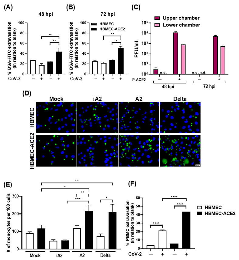
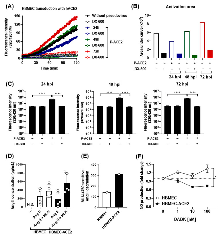
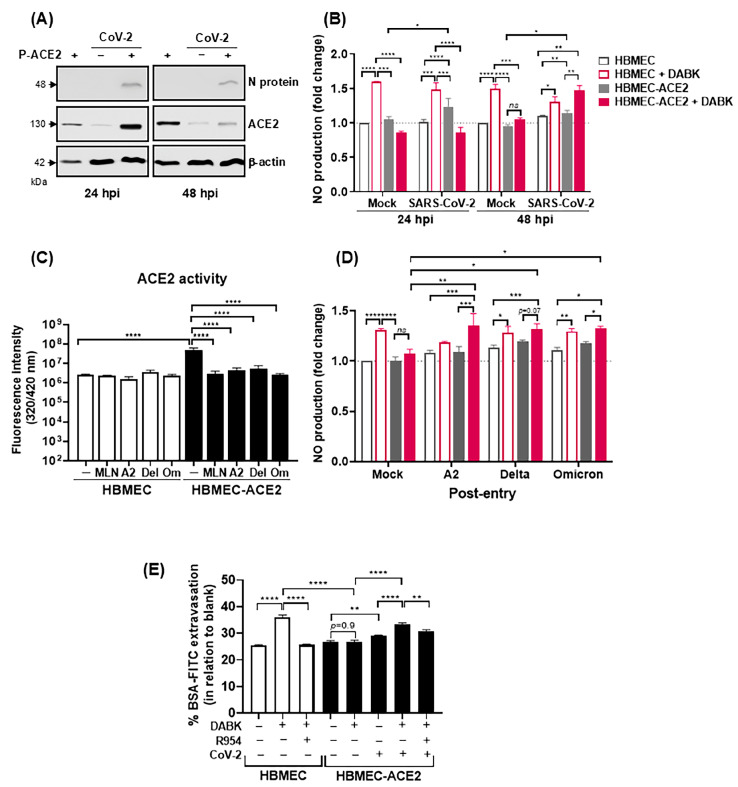
Endothelial dysfunction plays a central role in COVID-19 pathogenesis, by affecting vascular homeostasis and worsening thromboinflammation. This imbalance may contribute to blood-brain barrier (BBB) disruption, which has been reported in long COVID-19 patients with neurological sequelae. The kallikrein-kinin system (KKS) generates bradykinin (BK), a proinflammatory peptide that induces microvascular leakage via B2R. Under inflammatory conditions, BK is converted to Des-Arg-BK (DABK), which activates B1R, a receptor upregulated in inflamed tissues. DABK is degraded by ACE2, the main SARS-CoV-2 receptor; thus, viral binding and ACE2 downregulation may lead to DABK/B1R imbalance. Here, we investigated these interactions using human brain microvascular endothelial cells (HBMECs), as a model of the BBB. Since endothelial cell lines express low levels of ACE2, HBMECs were modified with an ACE2-carrying pseudovirus. SARS-CoV-2 replication was confirmed by RNA, protein expression, and infectious particles release. Infection upregulated cytokines and endothelial permeability, enhancing viral and leukocyte transmigration. Additionally, viral replication impaired ACE2 function in HBMECs, amplifying the response to DABK, increasing nitric oxide (NO) production, and further disrupting endothelial integrity. Our findings reveal a mechanism by which SARS-CoV-2 impacts the BBB and highlights the ACE2/KKS/B1R axis as a potential contributor to long COVID-19 neurological symptoms.

Keywords: ACE2; SARS-CoV-2; blood–brain barrier; des-Arg-bradykinin; endothelial cells; inflammation.


Conflict of interest statement

The authors declare no conflicts of interest.

Figures

Figure 1
Figure 1
Immortalized HBMECs do not sustain productive SARS-CoV-2 replication. (A) Expression of ACE2 mRNA in HBMECs and Vero-ACE2 was measured by RT-qPCR, and gapdh expression was used for normalization. Bars indicate the ratio between Ct GAPDH and ACE2. (BD) The expressions of ACE2 (B,C) and TMPRSS2 (D) were evaluated in HBMEC and Vero-ACE2 cells by Western blotting; relative expression in relation to β-actin was measured using ImageJ software version 8.0.2. (EH) HBMECs were inoculated with different SARS-CoV-2 lineages, using a MOI of 0.1. Cell lysates (E,F) and culture medium (G,H) were harvested at the indicated time points. Viral RNA (EG) and infectious virus particles (H) were measured by RT-qPCR and plaque assay. (I) Cells were infected as in (E) and cell viability was measured by LDH released in the culture medium. Data represent the average and SEM from three independent experiments; and were analyzed by unpaired t test. * p < 0.05; ** p < 0.005.
Figure 2
Figure 2
Development of HBMEC line overexpressing ACE2 by transient transduction with ACE2-carrying pseudovirus. (A) Schematic representation of the pseudovirus construction (P-ACE2). HEK-293 cells were transfected with pCMV-VSV-G, pHIV-1NL4-3 ΔEnv-NanoLuc, and pHAGE2-EF1a ACE2. After 24 h of transfection, the culture medium was replaced, the cells were cultured for another 24 h, and the supernatant containing pseudovirus was harvested. (B) HBMECs were transduced with different concentrations of P-ACE2 (based on p24 ELISA) and the cell viability was measured at the indicated time points by CellTiter Aqueous reagent. Data represent the average and SEM from three independent experiments; and were analyzed by one way ANOVA. ** p < 0.005; **** p < 0.0001. (C,D) HBMECs were transduced with P-ACE2 as in (B) and the expression of ACE2 was measured by Western blotting at the indicated time points. Representative Western blottings are demonstrated in (C) and the ratio between ACE2 and β-actin from 2 different assays is represented in (D).
Figure 3
Figure 3
HBMEC-ACE2 is permissive to SARS-CoV-2 replication. HBMECs were transduced or not with P-ACE2 for 48 h and then infected with different lineages of SARS-CoV-2. (A,B) Cell lysates were harvested at different time points post infection and the concentrations of genomic (gRNA) and subgenomic (sgRNA) virus RNA were measured by RT-qPCR. (C) Culture supernatants were harvested at the indicated periods and the released virus RNA was measured by RT-qPCR. (D) After 48 h, cells were incubated with J2 antibody and DAPI and the presence of dsRNA-positive cells was evaluated by fluorescence microscope. (E) SARS-CoV-2 N protein expression was measured by Western blotting at 24 and 48 hpi. (F,G) The titer of released infectious particles was measured by plaque assay. A representative assay is shown in (F) and the average and SEM from three different experiments are demonstrated in (G). Data were analyzed by two-way ANOVA. * p < 0.05; ** p < 0.005; *** p < 0.0005; **** p < 0.0001. (H) Cell viability was evaluated at different periods post infection by measurement of LDH released in the culture medium.
Figure 4
Figure 4
SARS-CoV-2 replication-dependent and -independent expression of inflammatory cytokines. HBMECs or HBMEC-ACE2 were inoculated with native (A2, Delta, Omicron) or inactivated (iA2, iDelta, iOmicron) SARS-CoV-2 from the lineages A2 (A), Delta (B), or Omicron BA1 (C). Cell lysates were harvested at 24 and 48 hpi, and the concentrations of mRNA corresponding to the indicated chemokines (CCL2, CCL5, IL-8) and cytokines (IL-6, TNF) were measured by RT-qPCR. Data represent the average and SEM from three independent experiments; and were analyzed by two-way ANOVA. * p < 0.05; ** p < 0.005; *** p < 0.0005; **** p < 0.0001. (D) After 24 hpi, cells were incubated with CM-H2DCFDA fluorescent probe, and the generation of reactive oxygen species (ROS) was evaluated by spectrophotometry; Heme was added as a positive control. (E) After 24 hpi, cells were incubated with DAF-FM diacetate fluorescent probe and nitric oxide (NO) production was evaluated by spectrophotometry; bradykinin (BK) was added to the cultures, as a positive control. The bars indicate the average and SEM from three independent experiments and data were analyzed by one way ANOVA, followed by Tukey’s multiple comparison test; * p < 0.05.
Figure 5
Figure 5
SARS-CoV-2 replication in HBMEC-ACE2 induces permeability, with crossing of viruses and mononuclear cells. HBMECs were transduced or not with P-ACE2 for 48 h and then infected with SARS-CoV-2. At 48 (A) and 72 hpi (B), the cells were incubated with BSA-FITC for 1 h and the amount of protein detected in the lower chamber was measured by fluorimetry. The bars represent the average and SEM from three independent experiments and the data were analyzed by unpaired t test. (C) At the indicated time points, culture medium from the upper and lower transwell chambers was harvested and the virus titers were measured by plaque assay. n.d.—not detected. (D,E) At 48 hpi, cells were incubated with CFSE-stained monocytes for 30 min and the number of adhered cells was analyzed by confocal fluorescence microscopy and measured using ImageJ software. Representative images are shown in (D) and the average and SEM from five independent experiments are indicated in (E). (F) HBMECs or HBMECs-ACE2 were cultured in a transwell system and, after 48 hpi, cells were incubated with PBMC. The percentage of PBMCs in the lower transwell chamber was calculated in relation to the wells with no cells (blank-100%). Data are representative of two independent assays. The data were analyzed by one way ANOVA, followed by Tukey’s multiple comparison test; * p < 0.05; ** p < 0.005; *** p < 0.0005; **** p < 0.0001.
Figure 6
Figure 6
ACE2 transduction in HBMECs downmodulates RAS and KKS. (A,B) Cell lysates obtained from HBMECs or HBMEC-ACE2 were incubated with the ACE2 fluorogenic substrate, in the presence or not of DX600. Activation of ACE2 was evaluated by measuring fluorescence emission due to substrate hydrolysis for 2 h. A representative curve obtained after spectrofluorimetric analysis is shown in (A), and the activation area, determined by subtracting the curve area obtained from lysates incubated or not with DX-600 is shown in (B). (C) After 24, 48, or 72 h post P-ACE2 transduction, intact culture cells were incubated with the ACE2 substrate for 2 h, and fluorescence emission was measured. (D,E) HBMEC or HBMEC-ACE2 were incubated with exogenous angiotensin II (AngII), in the presence or absence of MLN-4760. After 6 h, AngII concentration in the culture medium was measured by ELISA (D) and MLN-sensitive Ang II degradation (E) was calculated as the ratio between cultures with and without the ACE2 inhibitor. (F) HBMEC or HBMEC-ACE2 were incubated with increasing concentrations of DABK, and the production of nitric oxide (NO) was measured by spectrophotometry. The data were analyzed by one way ANOVA, followed by Tukey’s multiple comparison test; * p < 0.05; **** p < 0.0001; N.D.—not detected.
Figure 7
Figure 7
ACE2 downregulation by SARS-CoV-2 promotes an increase in DABK-mediated NO production and endothelial permeability. HBMECs were transduced or not with P-ACE2 for 48 h and then infected with SARS-CoV-2. (A) The expression of ACE2 was evaluated by Western blotting at 24 and 48 hpi. (B) After 24 or 48 hpi, the cells were incubated with des-Arg-BK (DABK) for 30 min and NO production was evaluated by spectrophotometry. (C) Cells were inoculated with the indicated SARS-CoV-2 lineages at an MOI of 10. At 1 h post entry, the fluorogenic ACE2 substrate was added, in the presence or not of MLN-4760. Enzymatic activity was measured by fluorescence emission in the culture medium. (D) Cells were infected as in (C) and then treated with DABK. NO production was measured as previously described. (E) HBMECs and HBMEC-ACE2 were cultured in a transwell system and infected or not with SARS-CoV-2 (delta lineage) for 48 h. Then, the cells were stimulated with DABK, in the presence or absence of R954. After 16 h stimulation, the cultures were incubated with BSA-FITC and permeability was measured by fluorimetry in the lower transwell chamber; data are representative of two independent experiments; the data were analyzed by one way ANOVA, followed by Tukey’s multiple comparison test; ns (not significant); * p < 0.05; ** p < 0.005; *** p < 0.0005; **** p < 0.0001.

Similar articles

References

    1. Huang C., Wang Y., Li X., Ren L., Zhao J., Hu Y., Zhang L., Fan G., Xu J., Gu X., et al. Clinical features of patients infected with 2019 novel coronavirus in Wuhan, China. Lancet. 2020;15:497–506. doi: 10.1016/S0140-6736(20)30183-5. - DOI - PMC - PubMed
    1. Zhou F., Yu T., Du R., Fan G., Liu Y., Liu Z., Xiang J., Wang Y., Song B., Gu X., et al. Clinical course and risk factors for mortality of adult inpatients with COVID-19 in Wuhan, China: A retrospective cohort study. Lancet. 2020;28:1054–1062. doi: 10.1016/S0140-6736(20)30566-3. - DOI - PMC - PubMed
    1. Lamers M.M., Haagmans B.L. SARS-CoV-2 pathogenesis. Nat. Rev. Microbiol. 2022;20:270–284. doi: 10.1038/s41579-022-00713-0. - DOI - PubMed
    1. Wang Y., Perlman S. COVID-19: Inflammatory Profile. Annu. Rev. Med. 2022;27:65–80. doi: 10.1146/annurev-med-042220-012417. - DOI - PubMed
    1. Mangalmurti N., Hunter C.A. Cytokine Storms: Understanding COVID-19. Immunity. 2020;14:19–25. doi: 10.1016/j.immuni.2020.06.017. - DOI - PMC - PubMed

MeSH terms

LinkOut - more resources