. 2025 Jun 12;26(12):5647.
doi: 10.3390/ijms26125647.

In Vitro Correction of Point Mutations in the DYSF Gene Using Prime Editing

Affiliations

In Vitro Correction of Point Mutations in the DYSF Gene Using Prime Editing

Camille Bouchard et al. Int J Mol Sci. .

Abstract

Dysferlinopathy is caused by over 500 mutations in the gene encoding dysferlin, including close to 300 point mutations. One option to cure the disease is to use a gene therapy to correct these mutations at the root. Prime editing is a technique which can replace the mutated nucleotide with the wild-type nucleotide. In this article, prime editing is used to correct several point mutations in the DYSF gene responsible for dysferlinopathy. In vitro editing of HEK293T cells reaches up to 31%. Notably, editing was more efficient in myoblasts than in patient-derived fibroblasts. The prime editing technique was also used to create a new myoblast clone containing a patient mutation from a healthy myoblast cell line.

Keywords: CRISPR; LGMD; Miyoshi Myopathy; dysferlin; dysferlinopathy; gene therapy; point mutation; prime editing.


Conflict of interest statement

The authors declare no conflicts of interest.

Figures

Figure 1
Figure 1
Percentage of editing in HEK293T by PE constructions made to correct the human DYSF E1833X and DYSF R1905X mutations. (A) Various pegRNA lengths designed to correct the DYSF E1833X mutation used in HEK293T cells: Different prime editing constructs made it possible to evaluate the optimal lengths of RTT and PBS to correct a given point mutation. For the E1833X mutation, this size was 13 nucleotides for both RTT and PBS. This construction made it possible to obtain 29% editing by transfection with Lipofectamine 2000 in HEK293T cells. (B) Comparing PE2 and PE3 using pegRNAs designed to correct the DYSF R1905X mutation used in HEK293T cells: Using an additional nicking single guide RNA (nsgRNA) to cut the DNA strand not modified by the epegRNA doubled the percentage of editing obtained, with epegRNA containing various RTT and PBS lengths. The figure illustrates PE2 (in light blue), using epegRNA to cut the DNA near the mutation, and PE3 constructs (in dark blue), using an additional nsgRNA, which are, on average, twice as effective at inserting a tracking mutation and allowed up to 31% of tracking mutations to be obtained in the HEK293T cells when the RTT contained 16 nucleotides and the PBS also contained 16 nucleotides. The experiment was repeated 3 times in 2 wells for each pegRNA construction.
Figure 2
Figure 2
Editing percentage in patient-derived fibroblasts. (A) DYSF R1905X: Editing in patient-derived fibroblasts has a maximum of 4% for the synonymous mutation added in the PAM and 5% for the correction of the patient’s R1905X mutation. The negative control with a plasmid containing the GFP gene indicates that 53% of the DNA does not have a pathological mutation, and the highest result is 58% for the RTT16-PBS13 construct (corresponding to 5% editing). (B) E1833X: The presence of E1833 increases by 20% (Ctrl = 55% E1833 vs. 75% E1833 for RTT16-PBS13), but the synonymous mutation of the PAM remains implanted at 0–4%.
Figure 3
Figure 3
Editing in patient-derived fibroblasts expressing MyoD. The patient-derived fibroblasts with the R1905X mutation were cultured with doxycycline to induce the expression of the lentivirus-inserted MyoD gene, and the patient fibroblasts were heterozygous (52% of the DYSF genes were wild-type on average when sequenced). The DNA after prime editing treatment was found to contain the wild-type DYSF gene in between 61% and 81% (red circle) (average 71%) of the cells. Thus, there was a 20% increase in the wild type genes. This means that 40% of the disease alleles had been corrected. The synonymous PAM mutation T is introduced at 1% (green circle) and the WT A (black circle) is present at 95%. The EditR Software colors the desired nucleotides from the sequence in blue and the undesired (background or spontaneous mutations) or voluntarily modified nucleotides in red as they differ from the given wild-type sequence. The experiment was repeated 3 times in 2 wells for each pegRNA construction.
Figure 4
Figure 4
Detection of potential off-target mutations. The online software Cas-OFFinder identifies genome sequences similar to those targeted by the spacer of the epegRNA. The constructs to correct the DYSF R1905X, E1833X and Q1010X mutations theoretically do not target any other sequence in the genome. However, the protospacer sequence near the W965X mutation is similar to a sequence in the ELAPOR2 gene (2 mismatches in the 20-nucleotide protospacer sequence) encoding the synthesis of a ubiquitous protein.
Figure 5
Figure 5
Two different protospacers may be used to insert the DYSF W965X mutation by prime editing. The position of the W965X (i.e., TGG (W)>TAG(Stop)) mutation in the DYSF gene is identified with a red square in bot (A,B). This figure illustrates that to insert this mutation, there are two possible PAMs and thus two possible protospacer sequences. Indeed, there is a TGG PAM in the bottom strand in Figure (A) and a TGG PAM in the top strand in Figure (B). In both cases, the protospacer sequences are composed of the 20 nucleotides located 5′ to the PAM. The break is performed at 3 nucleotides from the PAM in the 5′ direction. The cut site is the boundary between the RTT and the PBS. Their lengths are variable and specific to each sequence. However, only the PAM in Figure (A) can be used to correct the patient mutation, since the PAM in Figure (B) uses the wild type. Capital letters and their colour indicate the amino acid associated with the DNA sequence.
Figure 6
Figure 6
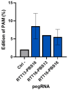
Percentage of insertion of the PAM synonymous mutation in healthy human myoblasts. A synonymous mutation was inserted in healthy human myoblasts in the PAM sequence used by 3 epegRNAs (with different RTT and PBS lengths). An average of 8.5% editing was obtained with the epegRNA containing an RTT with 13 nucleotides and a PBS with 16 nucleotides. The maximum insertion of the mutation was 11%. Each pegRNA was tested in two wells and the experiment was repeated a second time.
Figure 7
Figure 7
Insertion of the W965X mutation in healthy myoblasts. The human myoblasts cell line containing the W965X patient mutation is created by incorporating the mutation through electroporation of the plasmid of the prime editing components. The first electroporation makes it possible to obtain the mutation in 11% of the cells. The second treatment allows this figure to reach 25% (red circle). The EditR Software colors the desired nucleotides from the sequence in blue and the undesired (background or spontaneous mutations) or voluntarily modified nucleotides in red as they differ from the given wild-type sequence.
Figure 8
Figure 8
Description of CRISPR-Cas9 and prime editing. Colours in the text identify structures of the same colour in the figure above. The red X represents the patient mutation.
Figure 9
Figure 9
Construction and use of plasmids for prime editing. For the fabrication of prime editing plasmids, we used the protocol initially developed by Dr. David R. Liu’s team [26]. The first step was to digest the P2U6 plasmid (made from the pU6-tevopreq1-GG-acceptor (Addgene #174038), to which our laboratory added a second U6 promoter to insert the nsgRNA sequence) with the BsaI enzyme, removing the part with a red X and to insert the PBS and the RTT sequences between the sticky ends generated by the digestion. A transformation allowed for the isolation of a colony with the right sequence. The resulting plasmid (illustrated by a circle with arrows) was digested again with BbsI to add the sequence of a nicking single guide RNA (nsgRNA) to cut the DNA strand not modified by the epegRNA. A second transformation allowed for the selection of a bacterial colony containing the plasmid with the desired sequence. These plasmids were then used to treat different types of cells co-transfected with PE2-CMV (Addgene # 132775) by electroporation (Neonfection) or lipofection (Lipofectamine 2000, Thermo Fisher, Waltham, MA, USA). The DNA from the treated cells was extracted after 72 h, the DNA region near the targeted mutation (250–300 nucleotides) was PCR-amplified, and the amplicons were Sanger-sequenced and the sequence was analyzed with EditR software online (http://baseeditr.com/, last consulted on 7 June 2025). The colours used in the graph indicate the sequence nucleotides.

Similar articles

References

    1. Bouchard C., Tremblay J.P. Limb-girdle muscular dystrophies classification and therapies. J. Clin. Med. 2023;12:4769. doi: 10.3390/jcm12144769. - DOI - PMC - PubMed
    1. Straub V., Murphy A., Udd B., Corrado A., Aymé S., Bönneman C., de Visser M., Hamosh A., Jacobs L., Khizanishvili N. 229th ENMC international workshop: Limb girdle muscular dystrophies-Nomenclature and reformed classification Naarden, the Netherlands, 17–19 March 2017. Neuromuscul. Disord. 2018;28:702–710. doi: 10.1016/j.nmd.2018.05.007. - DOI - PubMed
    1. Bouchard C., Tremblay J.P. Portrait of dysferlinopathy: Diagnosis and development of therapy. J. Clin. Med. 2023;12:6011. doi: 10.3390/jcm12186011. - DOI - PMC - PubMed
    1. Liu J., Aoki M., Illa I., Wu C., Fardeau M., Angelini C., Serrano C., Urtizberea J.A., Hentati F., Hamida M.B. Dysferlin, a novel skeletal muscle gene, is mutated in Miyoshi myopathy and limb girdle muscular dystrophy. Nat. Genet. 1998;20:31–36. doi: 10.1038/1682. - DOI - PubMed
    1. Muriel J., Lukyanenko V., Kwiatkowski T., Bhattacharya S., Garman D., Weisleder N., Bloch R.J. The C2 domains of dysferlin: Roles in membrane localization, Ca2+ signalling and sarcolemmal repair. J. Physiol. 2022;600:1953–1968. doi: 10.1113/JP282648. - DOI - PMC - PubMed

Grants and funding

LinkOut - more resources