. 2025 Jun 13;26(12):5680.
doi: 10.3390/ijms26125680.

The Anti-Parkinsonian A2A Receptor Antagonist Istradefylline (KW-6002) Attenuates Behavioral Abnormalities, Neuroinflammation, and Neurodegeneration in Cerebral Ischemia: An Adenosinergic Signaling Link Between Stroke and Parkinson's Disease

Affiliations

The Anti-Parkinsonian A2A Receptor Antagonist Istradefylline (KW-6002) Attenuates Behavioral Abnormalities, Neuroinflammation, and Neurodegeneration in Cerebral Ischemia: An Adenosinergic Signaling Link Between Stroke and Parkinson's Disease

Michael G Zaki et al. Int J Mol Sci. .

Abstract

Stroke, the third leading cause of death worldwide, is a major cause of functional disability. Cerebral ischemia causes a rapid elevation of adenosine, the main neuromodulator in the brain. The inhibition of adenosine A2A receptors (A2ARs) has been introduced as a potential target in neurodegenerative disorders involving extracellular adenosine elevation. Istradefylline, a selective A2AR antagonist, has been approved for Parkinson's disease (PD) adjunctive therapy and showed neuroprotective effects in PD and Alzheimer's disease. However, the role of A2ARs in post-stroke neuronal damage and behavioral deficits remains unclear. We recently showed that A2AR antagonism prevented the adenosine-induced post-hypoxia synaptic potentiation of glutamatergic neurotransmission following the hypoxia/reperfusion of hippocampal slices. Here, we investigated the potential neuroprotective effects of istradefylline in male Sprague-Dawley rats subjected to pial vessel disruption (PVD) used to model a small-vessel stroke. Rats were treated with either a vehicle control or istradefylline (3 mg/kg i.p.) following PVD surgery for three days. Istradefylline administration prevented anxiety and depressive-like behaviors caused by PVD stroke. In addition, istradefylline significantly attenuated ischemia-induced cognitive impairment and motor deficits. Moreover, istradefylline markedly reduced hippocampal neurodegeneration, as well as GFAP/Iba-1, TNF-α, nNOS, and iNOS levels after PVD, but prevented the downregulation of anti-inflammatory markers TGF-β1 and IL-4. Together, these results suggest a molecular link between stroke and PD and that the anti-PD drug istradefylline displays translational potential for drug repurposing as a neuroprotective agent for cerebral ischemic damage.

Keywords: adenosine A1 receptor; adenosine A2A receptor; fEPSP; glutamate excitotoxicity; ischemic stroke; istradefylline; stroke model.


Conflict of interest statement

The authors have no relevant financial or non-financial interests to disclose.

Figures

Figure 1
Figure 1
Istradefylline decreased presynaptic glutamate release and prevented the adenosine-induced post-hypoxia synaptic potentiation (APSP) following hypoxia/reperfusion. Hippocampal slices were preincubated with 1 µM istradefylline for 1 h before hypoxia. (A) Sample traces of the field excitatory postsynaptic potential (fEPSP) experiment showing an average of the last 5 min of the 10 min baseline in black color (1), 20 min hypoxia in red color (2), 45 min normoxic washout in green color (3), and an overlay of all traces (1 + 2 + 3). Scale bars show 10 ms (x) and 0.5 mV (y). (B) Time course graph showing normalized mean fEPSP of both control (no istradefylline in black color) and slices preincubated with istradefylline (gray color). (C) Summary bar graph showing the average fEPSP value as a percentage of the baseline (100%) in the last 5 min of normoxic washout. Istradefylline prevented the development of APSP, whereas control slices showed facilitated fEPSPs during normoxia. (D) Bar chart showing the % PPR calculated in the last 5 min of normoxic washout and normalized to baseline. All graphed values showed mean ± SEM. N = 6 independent fEPSP recordings per treatment group; N = 6 independent experiments for PI fluorescence images. Significance: * = p < 0.05 and ** = p < 0.01.
Figure 2
Figure 2
Administration of istradefylline significantly attenuated hippocampal cell death caused by hypoxia. Hippocampal slices were pretreated with 1 µM istradefylline for 1 h prior to hypoxia and stained with propidium iodide (PI), a fluorescent label for cell death. (A) Full hippocampal slices fluorescently stained with PI, taken at 10× magnification, showing PI fluorescence. Increased PI fluorescence indicates increased cell death. (B) Representative PI fluorescence images of the CA1 hippocampal cell layer (white boxed region in (A)) taken at 63× magnification. Scale bars: 1 mm (whole hippocampus, in (A)) and 10 µm (CA1, in (B)). (C) Bar graph showing analyzed densitometry values of the zoomed CA1 images (shown in (B)) to compare relative PI fluorescence intensity between treatment groups (control with no hypoxia in black color, hypoxia treated with vehicle control in red color, and hypoxia treated with istradefylline in gray color). All values were normalized to control (100%). All values showed mean ± SEM. Significance: ** = p < 0.01 and *** = p < 0.001. N = 6 independent experiments.
Figure 3
Figure 3
Administration of istradefylline prevented cognitive impairments caused by focal cortical ischemia. The PVD group exhibited the least time spent exploring the novel arm and the highest percentage of time spent in the old arm compared to the other treatment groups. Arm durations were calculated as a percentage of the 5 min second retrieval trial. (AC) Bar graphs represent the percentage of time spent in novel, old, and start arms, respectively. (DF) Representative heat maps of the 5 min second trial acquired from Ethovision for sham, PVD + DMSO/saline, and PVD + istradefylline 3 mg/kg, respectively. N values = 12 for each treatment group. Values are shown as mean ± SEM. Significance values: *** = p < 0.001.
Figure 4
Figure 4
Istradefylline showed anxiolytic effects and inhibited motor deficits following ischemic stroke. Istradefylline significantly increased the percentage of time spent in the center square field and the number of entries into the center of the open field. In addition, A2AR antagonism restored motor activity and inhibited the reduction in distance moved by the rats in the maze during the task. (A) Bar chart showing the percentage of time spent in the center square of the field during the 15 min trial. (B) Bar chart showing the average number of entries into the center square of the maze during the task. (C) Bar chart showing the average distance (in cm) traveled by the rats in the field. (DF) Representative heat maps from each treatment group were acquired by EthoVision. (G) Rats were pre-trained on the rotarod for 2 days before induction (two trials each day) and then tested 72 h following PVD surgery. The bar chart shows the average maximum time spent on the rotarod before falling. Istradefylline-treated rats preserved the overall motor function; however, the PVD–vehicle control group had the least latency before falling from the rotarod. N = 12 for each treatment group. Values are shown as mean ± SEM. Significance values: * = p < 0.05, ** = p < 0.01, and *** = p < 0.001.
Figure 5
Figure 5
Istradefylline prevented depressive-like behavior. Rats were placed in a water tank for 10 min, and time spent immobile was calculated as a percentage of the total trial time. Latency to immobility was plotted as times in seconds. (A) The PVD–istradefylline-treated group attenuated the depressive-like behavior and showed a 50% reduction in time spent immobile compared to the vehicle control-treated group. (B) Istradefylline treatment showed 95% improvement in the latency time of immobility. (C,D) Average success and vigor scores were estimated as mentioned in the Methods Section. N = 12 for each treatment group. Values are shown as mean ± SEM. Significance values: * = p < 0.05, ** = p < 0.01, and *** = p < 0.001.
Figure 6
Figure 6
Istradefylline reduced hippocampal cell death caused by focal cortical ischemia. The administration of istradefylline (IST) showed neuroprotective properties and decreased hippocampal cell death in both ipsilateral and contralateral sides. Hippocampal slices were stained with propidium iodide (PI), a fluorescent marker for cell death. (A) Full montage of hippocampus showing PI fluorescence obtained with 10×. Increased PI fluorescence indicates increased cell death. (B) Magnification of 63× of the representative squares in A (white boxed region) showing the CA1 area of the hippocampus stained with PI, respectively. Scale bars: 1 mm (whole hippocampus, in (A)) and 10 µm (CA1, in (B)). (C) Summary bar graph showing relative PI intensity of the CA1 images shown in squares in (A) compared to the corresponding sham group. Levels of hippocampal neuronal damage were lower in the contralateral compared to the ipsilateral side of the PVD lesion. N = 5 for each treatment group. Values are shown as mean ± SEM. Significance values: * = p < 0.05, ** = p < 0.01, and *** = p < 0.001. NS = non-significant.
Figure 7
Figure 7
Istradefylline attenuated PVD-induced hippocampal neurodegeneration in both ipsilateral and contralateral sides. The administration of istradefylline attenuated neurodegeneration in the hippocampus following PVD. Coronal hippocampal slices were stained with FluoroJade C (FJC), a specific fluorescent marker for degenerating neurons. (A) Full montage of hippocampus fluorescently stained with FJC obtained with 10×. (B) Magnification of 63× of the representative squares in A (white boxed region) showing the CA1 area of the ipsilateral and contralateral hippocampus, respectively. Scale bars: 1 mm (whole hippocampus, in (A)) and 40 µm (CA1, in (B). (C) Summary bar graphs showing relative FJC intensity of the CA1 images shown in squares in (A). N = 5 for each treatment group. Values are shown as mean ± SEM. Significance values: * = p < 0.05 and ** = p < 0.01.
Figure 8
Figure 8
Istradefylline inhibited the activation of microglia and astrocytes. (A) Representative Western blot of hippocampal lysates showing expression levels of GFAP (marker for astrocytes), Iba-1 (marker for microglia), and beta-actin (loading control); N = 8 independent observations. (B) Summary bar charts showing a significant upregulation of GFAP and Iba-1 in PVD-treated rats and reversal by istradefylline. Values are expressed as mean ± SEM. Statistical significance was assessed using a one-way ANOVA test and a Tukey–Kramer multiple comparison test with a 95% confidence interval. Significance values: * = p < 0.05, ** = p < 0.01, and *** = p < 0.001. NS = non-significant.
Figure 9
Figure 9
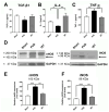
Istradefylline restored the balance between inflammatory and anti-inflammatory mediators following PVD. Treatment with istradefylline restored the levels of anti-inflammatory mediators TGF-β1 and IL-4 after PVD surgery (A,B). The PVD-induced elevation of inflammatory mediators TNF-α and iNOS (as well as nNOS) was attenuated with istradefylline treatment (CF). (AC) The total concentrations of TGF-β1, IL-4, and TNF-α were obtained using ELISA from ipsilateral hippocampal lysates. (D) Representative Western blot images of total ipsilateral hippocampal tissue lysates (left panel, nNOS and GAPDH; right panel, iNOS and GAPDH). (E,F) Summary bar charts showing relative expressions of nNOS (E) or iNOS (F) (% of sham). The administration of istradefylline prevented the PVD-induced increase in both nNOS (165 kDa monomeric nNOS; ~200 kDa believed to be nNOS bound to calmodulin) and iNOS (96 kDa iNOS cleavage product from the 130 kDa iNOS; see faint band above the 96 kDa signal). All tissue lysates were prepared 72 h following surgery. Values are shown as mean ± SEM. N = 5 in each group (independent samples). Significance values: * = p < 0.05, ** = p < 0.01, and *** = p < 0.01.
Figure 10
Figure 10
Schematic illustration of the ischemic stroke induction and treatment protocol to investigate the potential neuroprotective effects of istradefylline in a small-vessel-stroke model in male rats and in an ex vivo stroke model (hypoxia/reperfusion). Focal cortical ischemia was induced by pial blood vessel disruption (PVD) as described in the Methods Section. Rats subjected to PVD received either a vehicle control or istradefylline 1 h after the surgery. Behavioral tasks were performed on the third day to assess post-stroke memory deficits and other behavioral abnormalities. On the fourth day, the rats were sacrificed for subsequent post-mortem analysis of brain tissue samples.

Similar articles

References

    1. Hankey G.J. Stroke. Lancet. 2017;389:641–654. doi: 10.1016/S0140-6736(16)30962-X. - DOI - PubMed
    1. Lindsay M.P., Norrving B., Sacco R.L., Brainin M., Hacke W., Martins S., Pandian J., Feigin V. World Stroke Organization (WSO): Global Stroke Fact Sheet 2019. Int. J. Stroke Off. J. Int. Stroke Soc. 2019;14:806–817. doi: 10.1177/1747493019881353. - DOI - PubMed
    1. Benjamin E.J., Virani S.S., Callaway C.W., Chang A.R., Cheng S., Chiuve S.E., Cushman M., Delling F.N., Deo R., de Ferranti S.D., et al. Heart Disease and Stroke Statistics—2018 Update: A Report From the American Heart Association. Circulation. 2018;137:e67–e492. doi: 10.1161/CIR.0000000000000558. - DOI - PubMed
    1. Virani S.S., Alonso A., Benjamin E.J., Bittencourt M.S., Callaway C.W., Carson A.P., Chamberlain A.M., Chang A.R., Cheng S., Delling F.N., et al. Heart disease and stroke statistics—2020 update: A report from the American Heart Association. Circulation. 2020;141:e139–e596. doi: 10.1161/CIR.0000000000000757. - DOI - PubMed
    1. Katsanos A.H., Palaiodimou L., Zand R., Yaghi S., Kamel H., Navi B.B., Turc G., Romoli M., Sharma V., Mavridis D., et al. Abstract P82: The Impact of SARS-CoV-2 on Stroke Epidemiology and Care: A Meta-Analysis. Stroke. 2021;52((Suppl. 1)):AP82. doi: 10.1161/str.52.suppl_1.P82. - DOI - PMC - PubMed

MeSH terms

LinkOut - more resources