. 2025 Jun 13;26(12):5683.
doi: 10.3390/ijms26125683.

Anti-lymphoma Activity of Acyclic Terpenoids and Its Structure-Activity Relationship: In Vivo, In Vitro, and In Silico Studies

Affiliations

Anti-lymphoma Activity of Acyclic Terpenoids and Its Structure-Activity Relationship: In Vivo, In Vitro, and In Silico Studies

Fernando Calzada et al. Int J Mol Sci. .

Abstract

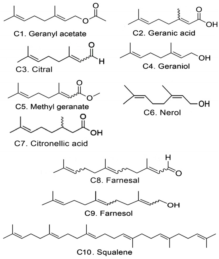
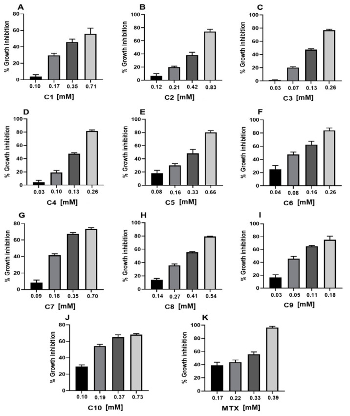
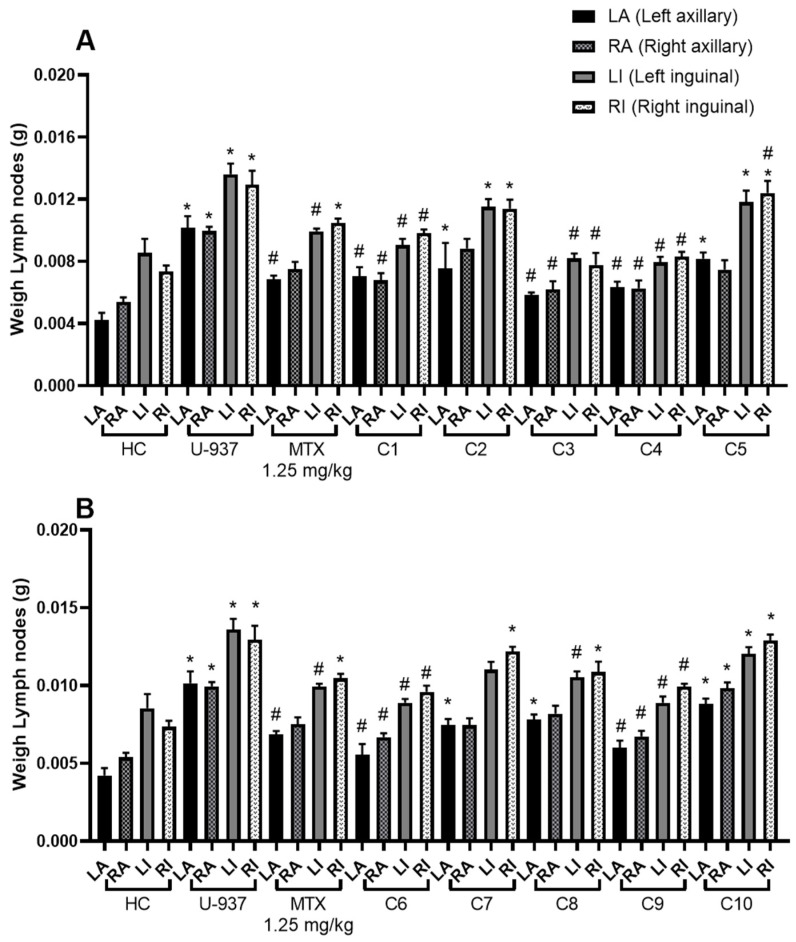
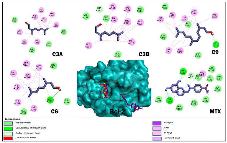
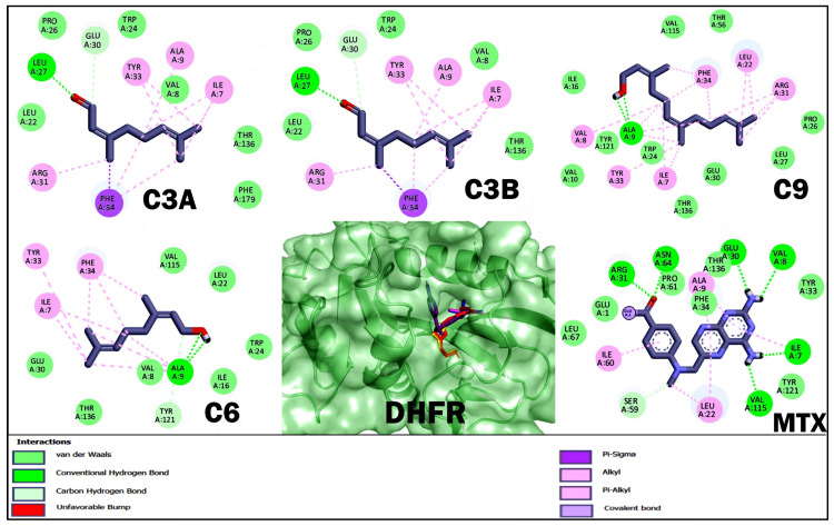
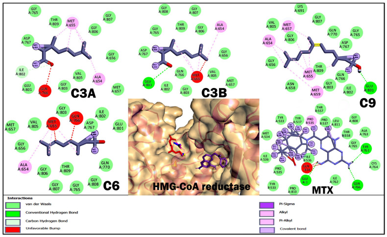
Terpenoids are a large group of molecules present in several plant species and in many essential oils reported with cytotoxic and anticancer properties. The aim of this study was to evaluate the anticancer activity of eleven acyclic terpenes; seven monoterpenoids: geranyl acetate (C1), geranic acid (C2), citral (C3, mixture of neral and geranial), geraniol (C4), methyl geranate (C5), nerol (C6) and citronellic acid (C7); two sesquiterpenes: farnesal (C8) and farnesol (C9); and one triterpene: squalene (C10), using in vivo, in vitro, and in silico models. Anti-lymphoma activity was evaluated using male Balb/c mice inoculated with U-937 cells. Cytotoxic activity was evaluated using the WST-1 method. Computer tools were used to obtain a molecular docking study, measuring pharmacokinetic and toxicological properties of the acyclic terpenoids with greater antitumor activity. The results showed that the terpenoids with the highest cytotoxic and nodal growth inhibitory activity were C3, C4, C6, and C9, and their effects were better compared to MTX. The data obtained suggest that the anti-lymphoma activity could be due to the presence of the aldehyde, hydroxyl, and acetate groups in the C1 of the monoterpenes and sesquiterpenes evaluated. The theoretical results obtained from molecular docking showed that geranial (C3A), neral (C3B), C9, and C6 terpenoids obtained a higher affinity for the HMG-CoA reductase enzyme and suggest that it could be a target to induce anti-lymphoma activity of bioactive terpenoids. Our study provides evidence that C3, C6, and C9 could be potential anticancer agents for the treatment of histiocytic lymphoma.

Keywords: Bcl-2; DHFR; FASN; HMG-CoA reductase; U-937 cells; acyclic terpenoids; anti-lymphoma activity; molecular docking.


Conflict of interest statement

The authors declare no conflicts of interest.

Figures

Figure 1
Figure 1
Structure of acyclic terpenoids proposed for the study.
Figure 2
Figure 2
Cytotoxic activity of acyclic terpenoids in U-937 cell line. Representative graphs show inhibition of cell growth caused by geranyl acetate (A), geranic acid (B), citral (C), geraniol (D), methyl geranate (E), nerol (F), citronellic acid (G), farnesal (H), farnesol (I), squalene (J), and MTX (K) at different concentrations after 24 h of exposure, (n = 3).
Figure 3
Figure 3
Lymph node weight (g) of male mice. The graph shows the weight of the axillary and inguinal lymph nodes as follows: left axillary (LA), right axillary (RA), left inguinal (LI), and right inguinal (RI). Healthy control (HC tween 80, 2% v/v in water), untreated control (U-937), methotrexate (MTX) 1.25 mg/kg. (A) Geranyl acetate (C1), geranic acid (C2), citral (C3), geraniol (C4), methyl geranate (C5); (B) nerol (C6), citronellic acid (C7), farnesal (C8), farnesol (C9), and squalene (C10) at 10 mg/kg. Results obtained by one-way ANOVA analysis followed by Dunnett’s test for multiple comparison. The data are expressed as mean ± SEM, (n = 6); * p < 0.05 vs. HC, # p < 0.05 vs. U-937.
Figure 4
Figure 4
Results of molecular docking of the enzyme Bcl-2 and terpenoids. The images show the position of the binding site and the 3D and 2D interactions. In the protein complex, the colors of terpenoids are shown as follows: geranial (C3A) magenta, neral (C3B) blue, nerol (C6) yellow, farnesol (C9) red, and methotrexate (MTX) in purple. In the 2D diagram, polar-type interactions are marked in green (van der waals, conventional hydrogen bond or carbon hydrogen bond); and non-polar-type interactions are marked in pink (alkyl or pi-alkyl), lilac (covalent bond), and purple (pi-sigma).
Figure 5
Figure 5
Results of molecular docking of the DHFR enzyme and terpenoids. The images show the position of the binding site and the 3D and 2D interactions. In the protein complex, the colors of terpenoids are shown as follows: geranial (C3A) magenta, neral (C3B) blue, nerol (C6) yellow, farnesol (C9) red, and methotrexate (MTX) in purple. In the 2D diagram, polar-type interactions are marked in green (van der waals, conventional hydrogen bond or carbon hydrogen bond); and non-polar-type interactions are marked in pink (alkyl or pi-alkyl), lilac (covalent bond), and purple (pi-sigma).
Figure 6
Figure 6
Results of molecular docking of the enzyme HMG-CoA reductase and terpenoids. The images show the position of the binding site and the 3D and 2D interactions. In the protein complex, the colors of terpenoids are shown as follows: geranial (C3A) magenta, neral (C3B) blue, nerol (C6) yellow, farnesol (C9) red, and methotrexate (MTX) in purple. In the 2D diagram, polar-type interactions are marked in green (van der waals, conventional hydrogen bond or carbon hydrogen bond); and non-polar-type interactions are marked in pink (alkyl or pi-alkyl), lilac (covalent bond), purple (pi-sigma) and red (unfavorable bump).
Figure 7
Figure 7
Results of the molecular docking of the FASN enzyme and terpenoids. The images show the position of the binding site and the 3D and 2D interactions. In the protein complex, the colors of terpenoids are shown as follows: geranial (C3A) magenta, neral (C3B) blue, nerol (C6) yellow, farnesol (C9) red, and methotrexate (MTX) in purple. In the 2D diagram, polar-type interactions are marked in green (van der waals, conventional hydrogen bond or carbon hydrogen bond); and non-polar-type interactions are marked in pink (alkyl or pi-alkyl), lilac (covalent bond), and purple (pi-sigma).

Similar articles

References

    1. Sun L., Romancik J.T. The Development and Application of Bispecific Antibodies in B-Cell Non-Hodgkin Lymphoma. J. Pers. Med. 2025;15:51. doi: 10.3390/jpm15020051. - DOI - PMC - PubMed
    1. Sapkota S., Shaikh H. Non-Hodgkin Lymphoma. StatPearls Publishing; Miami, FL, USA: 2023. StatPearls [Internet] - PubMed
    1. Luo J., Craver A., Bahl K., Stepniak L., Moore K., King J., Aschebrook-Kilfoy B. Etiology of non-Hodgkin lymphoma: A review from epidemiologic studies. J. Natl. Cancer Cent. 2022;2:226–234. doi: 10.1016/j.jncc.2022.08.003. - DOI - PMC - PubMed
    1. Secretaria de Salud Publica. [(accessed on 18 December 2024)]. Available online: https://www.gob.mx/salud/prensa/294-mexico-registra-al-ano-mas-de-195-mi....
    1. Leick M.B., Maus M.V., Frigault M.J. Clinical perspective: Treatment of aggressive B cell lymphomas with FDA-approved CAR-T cell therapies. Mol. Ther. 2021;29:433–441. doi: 10.1016/j.ymthe.2020.10.022. - DOI - PMC - PubMed

MeSH terms