Review
. 2025 Jun 15;26(12):5744.
doi: 10.3390/ijms26125744.

Ferritin in Acute Myeloid Leukemia: Not Only a Marker of Inflammation and Iron Overload, but Also a Regulator of Cellular Iron Metabolism, Signaling and Communication

Affiliations
Review

Ferritin in Acute Myeloid Leukemia: Not Only a Marker of Inflammation and Iron Overload, but Also a Regulator of Cellular Iron Metabolism, Signaling and Communication

Håkon Reikvam et al. Int J Mol Sci. .

Abstract

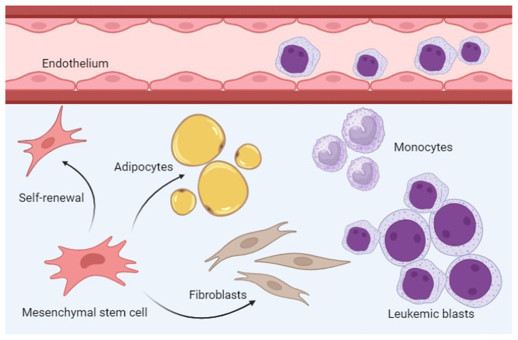
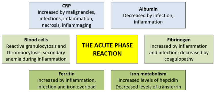
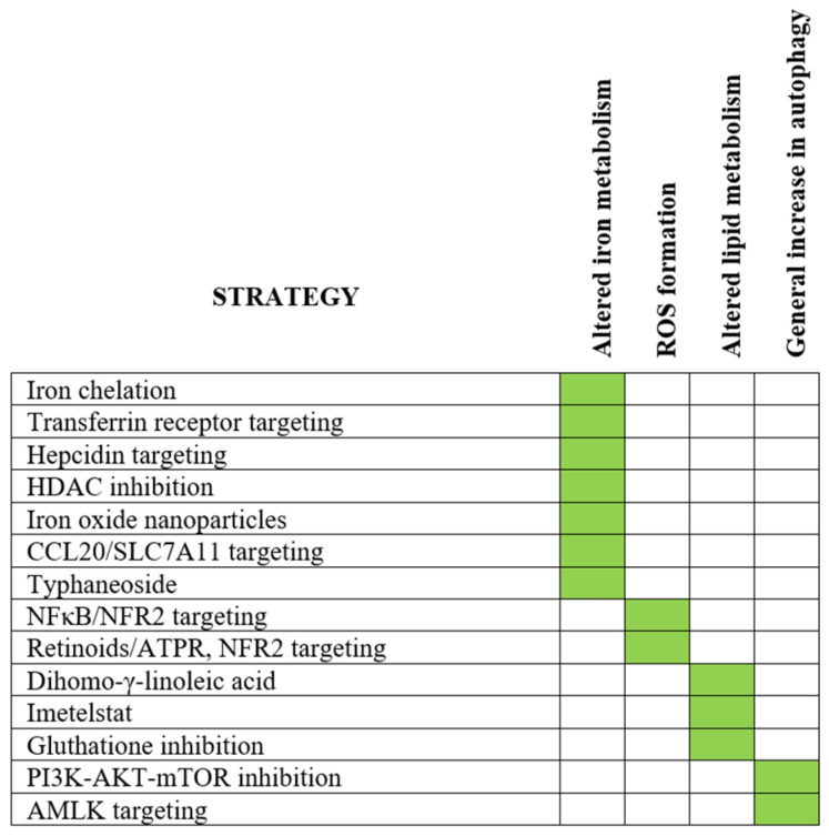
Ferritin is important for cellular iron storage and metabolism. It consists of 24 ferritin heavy- or light-chain subunits surrounding an iron-containing core, but it is also released as an extracellular molecule that shows increased systemic levels during acute-phase reactions. Furthermore, acute myeloid leukemia (AML) is an aggressive bone marrow malignancy that can be associated with increased ferritin levels both at the time of first diagnosis but also during/following anti-AML treatment due to an iron overload. Such high systemic ferritin levels at diagnosis or later allogeneic stem cell transplantation are associated with decreased long-term survival. Extracellular ferritin binds to several receptors expressed by AML cells (e.g., the transferrin receptor and CXCR4 chemokine receptor) and AML-supporting non-leukemic bone marrow cells (e.g., endothelial, mesenchymal or immunocompetent cells). Ferritin can thereby affect the AML cells directly as well as indirectly via AML-supporting neighboring cells. Finally, ferritin should be regarded as a regulator of the dysfunctional iron metabolism that causes increased iron levels in AML cells, and it is important for cell survival through its function during the initial steps of ferroptosis. Thus, ferritin is not only an adverse prognostic biomarker, but also an important regulator of AML cell proliferation, survival and chemosensitivity and the targeting of iron metabolism/ferroptosis is, therefore, a possible strategy in AML therapy.

Keywords: acute myeloid leukemia; acute-phase reaction; chemotherapy; ferritin; ferroptosis; iron; prognosis; stem cell transplantation.


Conflict of interest statement

The authors declare no conflicts of interest.

Figures

Figure 1
Figure 1
The bone marrow microenvironment in human AML; non-leukemic cells that (i) communicate with hierarchically organized AML cells and (ii) are influenced by soluble extracellular ferritin. Transendothelial AML cell migration with peripheral blood leukemization is seen for many patients, whereas extramedullary infiltration is uncommon. Soluble extracellular ferritin can affect at least endothelial cells, mesenchymal stem cells, fibroblast subsets and adipocytes as well as monocytes/macrophages. Possible ferritin effects on other AML-supporting cells (see the text) have not been characterized, and these cells are not shown in the figure.
Figure 2
Figure 2
The effects of the acute-phase reaction on iron metabolism are complex; ferritin levels increase whereas, at the same time, transferrin levels are decreased [110]. The serum levels of iron are, in addition, decreased and this seems to be at least partly due to the accumulation of iron in macrophages [110]. Furthermore, the effect of inflammation/an acute-phase reaction on the levels of circulating normal myeloid cells are divergent, neutrophils are increased, thrombocytosis is also common, and many patients have anemia [110]. Finally, the function of the hepatocytes that express/release many of the acute-phase proteins is altered and there are additional systemic neuroendocrine changes (e.g., increased catecholamines, corticotropin and cortisone), several metabolic changes (e.g., a loss of muscle and negative nitrogen balance, increased vasopressin, decreased IGF-1, increased hepatic lipogenesis, but increased lipolysis in adipose tissue) and changes in not-protein plasma constituents (e.g., hypozinchemia, hypercupremia and increased glutathione) [110,111]. The figure presents protein biomarkers of inflammation (blue), normal peripheral blood cells (including platelets)/coagulation (light green) and biomarkers of iron metabolism (dark green).
Figure 3
Figure 3
An overview of possible therapeutic strategies for targeting iron metabolism and/or induction of ferroptosis. The strategies are described more in detail in the text. Induction of ferroptosis includes the three main events: (i) altered iron metabolism, (ii) induction of oxidative stress and (iii) altered lipid metabolism (see Table 8) and, in addition, we indicate whether the main mechanism was a general increase in autophagy. The figure shows which of these three events that represent the main target for each of the described strategies (for references, please see the text in Section 9).

Similar articles

References

    1. Arber D.A., Orazi A., Hasserjian R.P., Borowitz M.J., Calvo K.R., Kvasnicka H.M., Wang S.A., Bagg A., Barbui T., Branford S., et al. International Consensus Classification of Myeloid Neoplasms and Acute Leukemias: Integrating morphologic, clinical, and genomic data. Blood. 2022;140:1200–1228. doi: 10.1182/blood.2022015850. - DOI - PMC - PubMed
    1. Salem M., Delwel R., Touw I., Mahmoud L., Löwenberg B. Human AML colony growth in serum-free culture. Leuk. Res. 1988;12:157–165. doi: 10.1016/0145-2126(88)90076-8. - DOI - PubMed
    1. Bruserud Ø., Frostad S., Foss B. In vitro culture of acute myelogenous leukemia blasts: A comparison of four different culture media. J. Hematother. 1999;8:63–73. doi: 10.1089/106161299320587. - DOI - PubMed
    1. Brenner A.K., Tvedt T.H., Nepstad I., Rye K.P., Hagen K.M., Reikvam H., Bruserud Ø. Patients with acute myeloid leukemia can be subclassified based on the constitutive cytokine release of the leukemic cells; the possible clinical relevance and the importance of cellular iron metabolism. Expert Opin. Ther. Targets. 2017;21:357–369. doi: 10.1080/14728222.2017.1300255. - DOI - PubMed
    1. Plays M., Müller S., Rodriguez R. Chemistry and biology of ferritin. Metallomics. 2021;13:mfab021. doi: 10.1093/mtomcs/mfab021. - DOI - PMC - PubMed

LinkOut - more resources