. 2025 Jun 16;26(12):5753.
doi: 10.3390/ijms26125753.

The SGLT2 Inhibitor Empagliflozin Mitigates the Harmful Effects of Methylglyoxal Exposure on Ovalbumin-Induced Mouse Airway Inflammation

Affiliations

The SGLT2 Inhibitor Empagliflozin Mitigates the Harmful Effects of Methylglyoxal Exposure on Ovalbumin-Induced Mouse Airway Inflammation

Matheus L Medeiros et al. Int J Mol Sci. .

Abstract

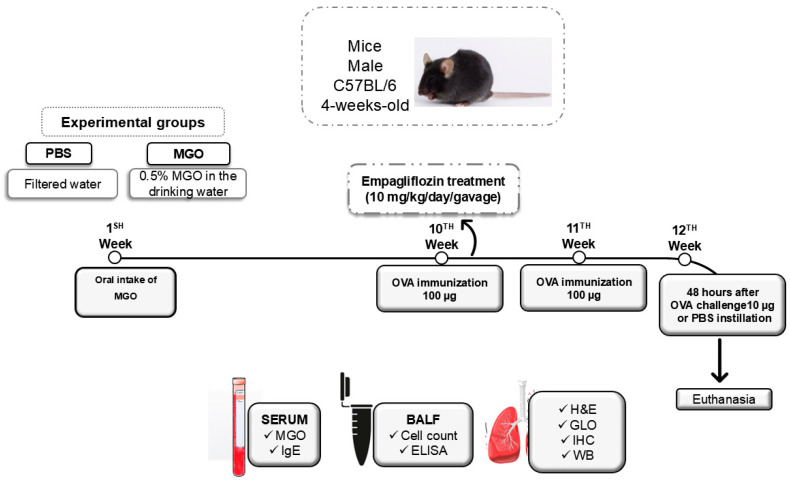
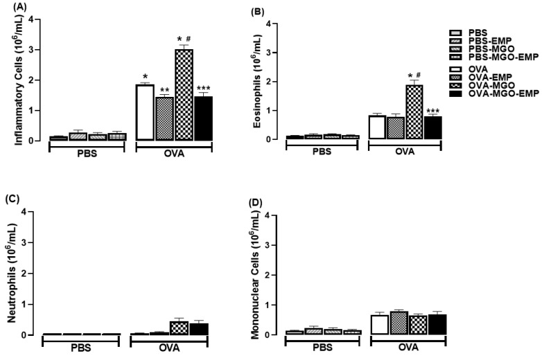
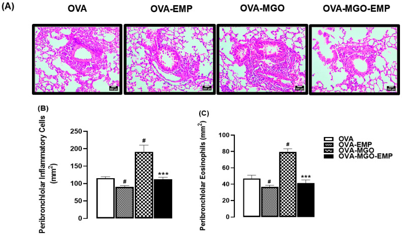
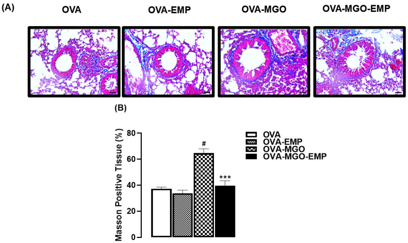
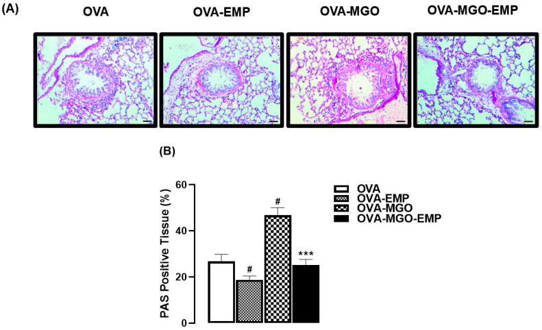
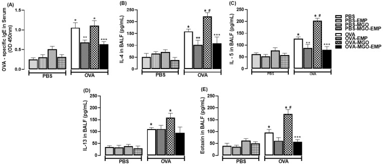
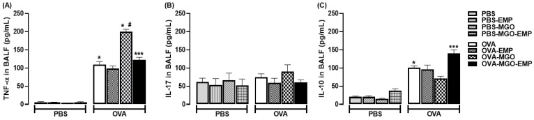
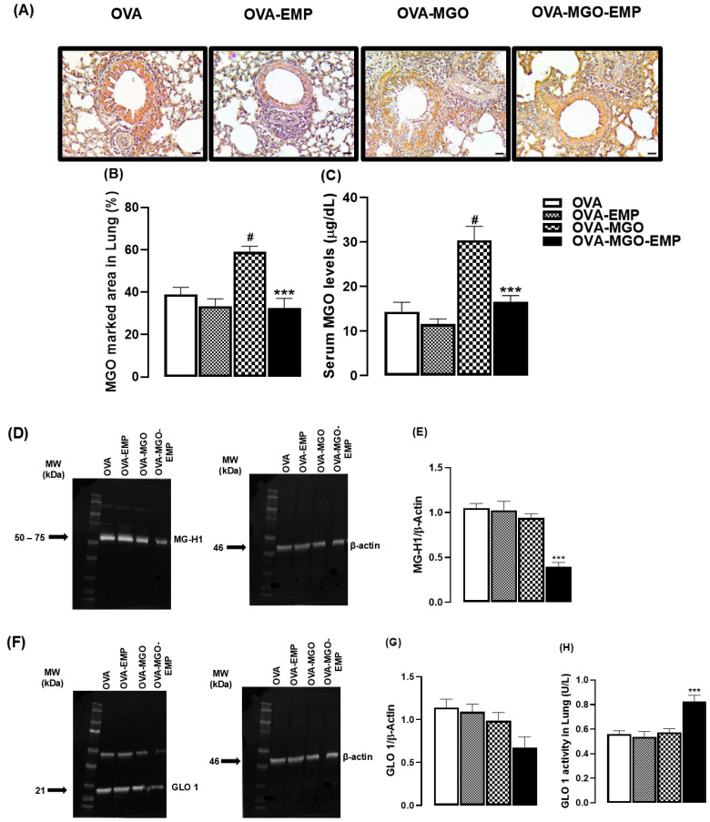
Asthma is a chronic inflammatory airway disease that can be aggravated by metabolic comorbidities such as type 2 diabetes mellitus (DM2) and obesity. Elevated levels of methylglyoxal (MGO), a reactive glycolysis byproduct, have been associated with exacerbation of allergic airway disease. SGLT2 inhibitors have been successfully employed in DM2 treatment. Here, we hypothesized that elimination of MGO might be a potential anti-inflammatory mechanism of SGLT2 inhibitors. This study aimed to evaluate the effects of empagliflozin on ovalbumin (OVA)-induced airway inflammation in mice chronically exposed to MGO. Male C57BL/6 mice sensitized with OVA were exposed to 0.5% MGO for 12 weeks and treated with empagliflozin (10 mg/kg, gavage, two weeks). MGO exposure significantly enhanced airway eosinophil infiltration, mucus production and collagen deposition, as well as levels of IL-4, IL-5, eotaxin and TNF-α. Empagliflozin treatment significantly reduced OVA-induced airway disease, which was accompanied by reductions in IgE, IL-4, IL-5, eotaxin, and TNF-α levels. Empagliflozin significantly reduced the MGO levels in serum, and immunohistochemical staining, and protein expression of MGO-hydroimidazolone (MG-H1), while increasing IL-10 levels and glyoxylase-1 (GLO 1) activity in lungs. In conclusion, empagliflozin efficiently removes MGO from circulation, while increasing the MGO detoxification by GLO 1, thereby mitigating the OVA-induced inflammation in MGO-exposed mice.

Keywords: MG-H1; asthma; collagen; cytokines; glyoxalase; mucus.


Conflict of interest statement

The authors declare no conflicts of interest.

Figures

Figure 1
Figure 1
Experimental protocol for immunization and challenge with ovalbumin (OVA): Male C57BL/6 mice were administered oral 0.5% methylglyoxal (MGO) for 12 weeks, with or without subsequent treatment with SGLT2 inhibitors during the final 2 weeks. Certain elements of the figure were created using images from Servier Medical Art, which is licensed under a Creative Commons Attribution 3.0 Unported License (https://creativecommons.org/licenses/by/3.0/ accessed on 10 April 2025). SGLT2, selective sodium-glucose cotransporter 2; BALF, bronchoalveolar lavage fluid; H&C, hematoxylin/eosin; IHC, immunohistochemistry; WB, Western blotting; GLO, glyoxalase.
Figure 2
Figure 2
Counts of total inflammatory cells (A), eosinophils (B), neutrophils (C), and mononuclear cells (D) in the bronchoalveolar lavage fluid (BALF) of mice intranasally challenged with ovalbumin (OVA). Mice were treated with 0.5% methylglyoxal (MGO) in drinking water for 12 weeks, either alone or in combination with empagliflozin (EMP). The data are expressed as mean ± standard error of the mean (SEM) with n = 5. * p < 0.05 compared to respective groups in PBS-instilled mice; ** p < 0.05 compared to respective OVA-EMP groups; # p < 0.05 compared to respective OVA groups; *** p < 0.05 compared to OVA-MGO groups.
Figure 3
Figure 3
Representative images of hematoxylin and eosin (H&E) staining (A), quantification of total inflammatory cells (B), and eosinophils (C) in lung tissue sections from ovalbumin (OVA)-challenged mice. Mice were treated with 0.5% methylglyoxal (MGO) in drinking water for 12 weeks, either alone or in combination with empagliflozin (EMP). The data are expressed as mean ± standard error of the mean (SEM) with n = 5. # p < 0.05 compared to respective OVA groups; *** p < 0.05 compared to the OVA-MGO groups. In panel A, scale bar = 200 μm (200× magnification).
Figure 4
Figure 4
Representative images (A) and quantification (%) of collagen deposition (B) in lung sections from ovalbumin (OVA)-challenged mice, as assessed by Masson’s trichrome staining. Mice were treated with 0.5% methylglyoxal (MGO) in drinking water for 12 weeks, either alone or in combination with empagliflozin (EMP). The data are expressed as mean ± standard error of the mean (SEM) with n = 5. # p < 0.05 compared to respective OVA groups; *** p < 0.05 compared to the MGO-OVA groups.
Figure 5
Figure 5
Representative images (A) and quantification (%) of mucus production (B) in lung sections from ovalbumin (OVA)-challenged mice, as assessed by periodic Acid-Schiff (PAS) staining. Mice were treated with 0.5% methylglyoxal (MGO) in drinking water for 12 weeks, either alone or in combination with empagliflozin (EMP). The data are expressed as mean ± standard error of the mean (SEM) with n = 5. # p < 0.05 compared to respective OVA groups; *** p < 0.05 compared to OVA-MGO group.
Figure 6
Figure 6
Levels of immunoglobulin IgE in serum (A) and IL-4 (B), IL-5 (C), IL-13 (D), and eotaxin (E) in bronchoalveolar lavage fluid (BALF) of mice instilled with phosphate-buffered saline (PBS) or intranasally challenged with ovalbumin (OVA). Mice were treated or not with 0.5% methylglyoxal (MGO) in drinking water for 12 weeks, either alone or in combination with empagliflozin (EMP). The data are expressed as mean ± standard error of the mean (SEM) with n = 5. * p < 0.05 compared to respective groups in PBS-instilled mice; ** p < 0.05 compared to respective OVA-EMP groups; # p < 0.05 compared to respective OVA groups; *** p < 0.05 compared to OVA-MGO groups.
Figure 7
Figure 7
Levels of TNF-α (A), IL-17 (B) and IL-10 (C) in bronchoalveolar lavage fluid (BALF) of mice instilled with phosphate-buffered saline (PBS) or intranasally challenged with ovalbumin (OVA). Mice were treated or not with 0.5% methylglyoxal (MGO) in drinking water for 12 weeks, either alone or in combination with empagliflozin (EMP). The data are expressed as mean ± standard error of the mean (SEM) with n = 5. * p < 0.05 compared to respective groups in PBS-instilled mice; # p < 0.05 compared to OVA group; *** p < 0.05 compared to OVA-MGO groups.
Figure 8
Figure 8
Immunohistochemical analysis of MG-H1 in lung tissue and methylglyoxal (MGO) levels in serum of mice treated with 0.5% MGO in drinking water for 12 weeks, either alone or in combination with empagliflozin (EMP). Representative immunohistochemistry images showing MG-H1 staining in lung tissue are shown in (A) with quantification of MG-H1-positive area (%) in lung sections (B). Panel (C) shows the serum levels of MGO. Panel (D) shows the Western blot analysis of MG-H1 expression in lung tissue (D) with corresponding densitometric quantification in (E). Panel (F) shows Western blot analysis of glyoxalase 1 (GLO 1) expression in lung tissue with densitometric quantification in (G). Enzymatic activity of GLO 1 in lung tissue is shown in (H). The data are expressed as mean ± standard error of the mean (SEM) with n = 5. # p < 0.05 compared to OVA group; *** p < 0.05 compared to OVA-MGO groups.

Similar articles

References

    1. Gyawali B., Georas S.N., Khurana S. Biologics in severe asthma: A state-of-the-art review. Eur Respir Rev. 2025;8:240088. doi: 10.1183/16000617.0088-2024. - DOI - PMC - PubMed
    1. Hussain M., Liu G. Eosinophilic Asthma: Pathophysiology and Therapeutic Horizons. Cells. 2024;13:384. doi: 10.3390/cells13050384. - DOI - PMC - PubMed
    1. Kroegel C., Virchow J.C., Luttmann W., Walker C., Warner J.A. Pulmonary immune cells in health and disease: The eosinophil leucocyte (Part I) Eur. Respir. J. 1994;7:519–543. doi: 10.1183/09031936.94.07030519. - DOI - PubMed
    1. Kuruvilla M.E., Lee F.E., Lee G.B. Understanding Asthma Phenotypes, Endotypes, and Mechanisms of Disease. Clin. Rev. Allergy Immunol. 2019;56:219–233. doi: 10.1007/s12016-018-8712-1. - DOI - PMC - PubMed
    1. Galli S.J., Tsai M. IgE and mast cells in allergic disease. Nat. Med. 2012;18:693–704. doi: 10.1038/nm.2755. - DOI - PMC - PubMed

MeSH terms