. 2025 Jun 16;26(12):5765.
doi: 10.3390/ijms26125765.

Molecular Insight into the Recognition of DNA by the DndCDE Complex in DNA Phosphorothioation

Affiliations

Molecular Insight into the Recognition of DNA by the DndCDE Complex in DNA Phosphorothioation

Wencheng Fu et al. Int J Mol Sci. .

Abstract

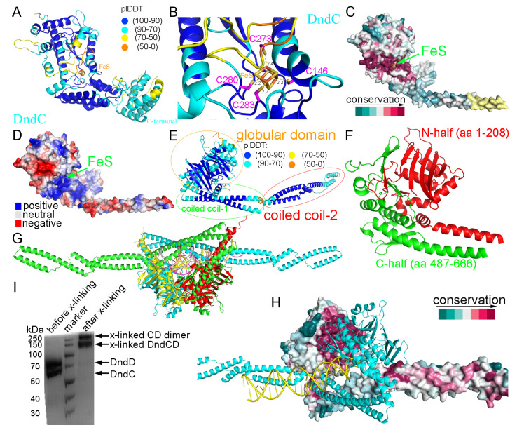
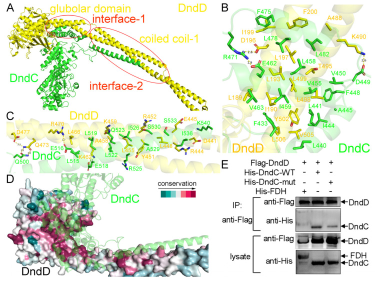
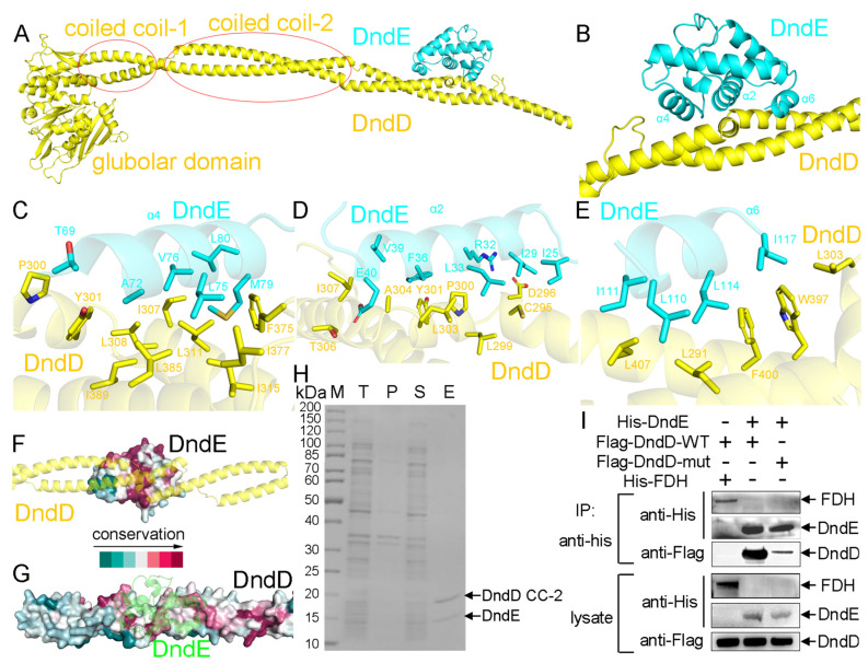
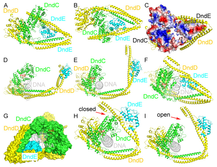
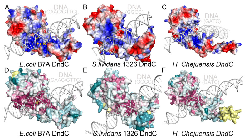
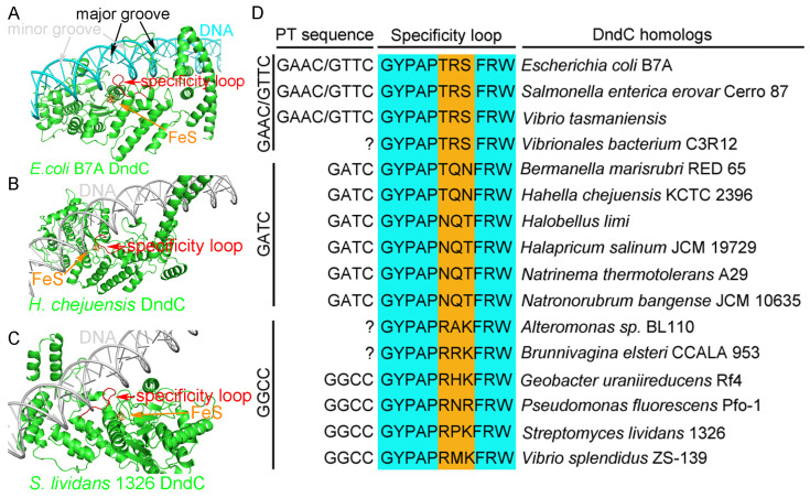
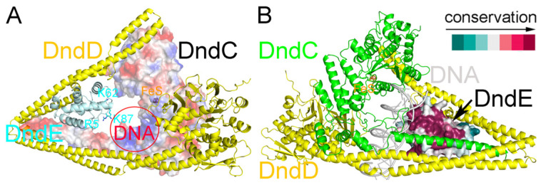
In a vast variety of prokaryotes such as Escherichia coli and Streptomyces lividans, the DNA degradation (Dnd) CDE protein complex (consisting of DndC, DndD, and DndE), together with the DndA/IscS protein and the DndFGH complex, function as a defense barrier to prevent the invasion of non-self-DNA. The DndCDE complex introduces phosphorothioation (PT) modifications into DNA, and the DndFGH complex specifically cleaves non-PT DNA and, thus, restricts horizontal gene transfer and phage invasion. Despite the central importance of the DndCDE complex in DNA PT modification, which catalyzes the oxygen-sulfur swap on DNA, our understanding of this key complex remains poor. Here, we employed protein structure prediction to provide a reasonably reliable prediction of the structure of the DndCDE complex and a 23 bp DNA-DndCDE complex. We found that among the three proteins in the DndCDE complex, DndC, especially its "specificity loop", plays a key role in recognizing the consensus PT modification sequence. In addition, the DndD protein is found to possess a highly conserved structural surface on its globular domain, presumably mediating the dimerization of DndD as well as the DndCDE complex. Furthermore, our normal mode analysis showed that there exists a dynamic transition between a closed and an open state for the DndCDE complex, facilitating its association and release of DNA. Our conclusions were corroborated by biochemical assays using purified proteins. On the whole, we provide molecular insights into the assembly and DNA-recognition mechanism of a central protein complex involved in DNA phosphorothioation.

Keywords: DNA-DndCDE complex; DndC; DndCDE complex; phosphorothioation modification; specificity loop.


Conflict of interest statement

The authors declare no conflicts of interest.

Figures

Figure 1
Figure 1
Predicted structures of DndC and DndD. (A) Predicted structure of E. coli B7A DndC. (B) The iron–sulfur cluster and the four conserved cysteine residues coordinating the iron–sulfur cluster. (C) The conservation of DndC across different species of prokaryotes was mapped onto the surface of its structure. The green arrow indicates the iron–sulfur cluster. (D) The electrostatic surface potential of DndC. (E) Predicted structure of E. coli B7A DndD. (F) Structure of the globular domain of DndD. (G) The AlphaFold3-predicted structure of the DndD dimer was structurally aligned with the Rad50 (red and yellow)-DNA (yellow and magenta) crystal structure using molecular superimposition. (H) The dimerization interface of DndD is highly conserved. (I) Cross-linking experiments using purified proteins of the DndC-DndD complex.
Figure 2
Figure 2
Predicted structure of the DndC-DndD complex. (A) Predicted structure of the DndC-DndD complex. There are two interaction interfaces between DndC and DndD, which are highlighted by red boxes in the figure. (B) In the first interface, the globular domain of DndD recognizes residues 433–482 of DndC. (C) In the second interface, the coiled coil-1 domain of DndD interacts with the very C-terminal α-helix of DndC. (D) Both of these two recognition surfaces are highly conserved across various species. (E) The nickel column pull-down assay using purified proteins of DndC and DndD.
Figure 3
Figure 3
DndD employs its coiled coil-2 domain to recognize DndE. (A) Predicted structure of the DndD-DndE complex. (B) DndE employs its α2, α4, and α6 helices to interact with DndD. (C) The binding interface between DndE-α4 and DndD. (D) The binding interface between DndE-α2 and DndD. (E) The binding interface between DndE-α6 and DndD. (F) The DndD-binding surface on DndE is highly conserved. (G) The DndE-binding surface on DndD is conserved across various species. (H) A pull-down experiment confirmed that the coiled coil-2 domain of DndD was sufficient for interacting with DndE. (I) Point mutations on the DndE-binding residues of DndD abolished its interaction with DndE.
Figure 4
Figure 4
Recognition of DNA by the DndCDE complex. (A) Predicted structure of the DndCDE complex. (B) Predicted structure of the DndCDE complex viewed from a different angle. (C) DndC and DndE provide the inner positively charged surface of the ring-like structure of the DndCDE complex, and DndD is located at the outside of the ring. (D) Side view of the predicted structure of E. coli B7A DndCDE in a complex with 23 bp of DNA (Supplementary Tables S1 and S2). (E) Top view of the predicted DndCDE-DNA complex structure (Supplementary Tables S1 and S2). (F) Oblique view of the predicted DndCDE-DNA complex structure (Supplementary Tables S1 and S2). (G) The cylindrical shape of DNA is complementary to that of the hollow cavity of the predicted DndCDE complex, with DndC and DndE contacting DNA (Supplementary Tables S1 and S2). A normal mode analysis confirmed that conformation transition enables the predicted DndCDE complex to cycle between association (H) with and dissociation (I) from DNA. The red arrows indicate the closed state (H) where the complex is associated with DNA, and the open state (I) where it is dissociated from DNA.
Figure 5
Figure 5
DndC is predicted to employ a highly conserved, positively charged groove for DNA recognition. (AC) DNA with GAAC/GTTC, GGCC/GGCC, and GATC/GATC repeat sequences are predicted to bind within the positively charged groove of E. coli B7A (A), S. lividans 1326 (B), and H. Chejuensis KCTC2396 DndC (C), respectively. (DF) A computational analysis predicts that the surfaces of the DNA-binding grooves of E. coli B7A (D), S. lividans 1326 (E), and H. Chejuensis KCTC2396 DndC (F) are highly conserved.
Figure 6
Figure 6
The specificity loop of DndC is predicted to recognize the core consensus phosphorothioation motif on DNA. The black arrow points to the DNA’s major groove, the gray arrow indicates the minor groove, the red arrow highlights the specificity loop, and the orange arrow marks the iron–sulfur cluster. (A) The specificity loop of E. coli B7A DndC is predicted to insert into the major groove of DNA and interact with the GAAC/GTTC core consensus phosphorothioation motif. (B) The specificity loop of H. chejuensis KCTC2396 DndC is predicted to insert into the major groove of DNA with the GATC sequence motif. (C) The specificity loop of S. lividans 1326 DndC is predicted to insert into the major groove of DNA with the GGCC sequence motif. (D) Three positions on the conserved specificity loop display subtype-specific conservation with respect to the three kinds of DNA PT modification. “?”indicates that the PT modification status is yet unknown.
Figure 7
Figure 7
Structural modeling predicts DNA recognition by DndE. (A) Three positively charged residues of E. coli B7A DndE are predicted to point toward the hollow space where the bound DNA might be located. (B) The predicted DNA-binding surface of DndE is highly conserved.
Figure 8
Figure 8
A conjugative transfer and an LC-LS assay supported the key role of the specificity loop of DndC in DNA PT. (A) The conjugative transfer between Streptomyces coelicolor M145 and E. coli with pHZ1904 plasmid did not occur (the left dish). The M145::pSET152 exconjugants and M145::pHZ1904-DndCTRS exconjugants grew normally (the middle and right dishes, respectively). (BD) Using the LC-MS assay to examine the GPTG (B), GPTA (C), and GPTT (D) modification, which comes from standard, pHZ1904, M145::pSET152 exconjugants, and M145::pHZ1904-DndCTRS exconjugants. For positive control, the GPTG, GPTA, and GPTT modification peaks from the standard and the GPTG modification peak from pHZ1904 plasmid were observed. However, GPTG, GPTA, and GPTT modification peaks from M145::pSET152 exconjugants and M145::pHZ1904-DndCTRS exconjugants were not detected. (M145 means Streptomyces coelicolor M145.) The dotted lines indicate the peak positions of the standard samples.

Similar articles

References

    1. Wang L., Jiang S., Deng Z., Dedon P.C., Chen S. DNA phosphorothioate modification–A new multi-functional epigenetic system in bacteria. FEMS Microbiol. Rev. 2019;43:109–122. doi: 10.1093/femsre/fuy036. - DOI - PMC - PubMed
    1. Ahlgren N.A., Chen Y., Needham D.M., Parada A.E., Sachdeva R., Trinh V., Chen T., Fuhrman J.A. Genome and epigenome of a novel marine Thaumarchaeota strain suggest viral infection, phosphorothioation DNA modification and multiple restriction systems. Environ. Microbiol. 2017;19:2434–2452. doi: 10.1111/1462-2920.13768. - DOI - PubMed
    1. Barbier P., Lunazzi A., Fujiwara-Nagata E., Avendaño-Herrera R., Bernardet J.F., Touchon M., Duchaud E. From the Flavobacterium genus to the phylum Bacteroidetes: Genomic analysis of dnd gene clusters. FEMS Microbiol. Lett. 2013;348:26–35. doi: 10.1111/1574-6968.12239. - DOI - PubMed
    1. Liu G., Fu W., Zhang Z., He Y., Yu H., Wang Y., Wang X., Zhao Y.L., Deng Z., Wu G., et al. Structural basis for the recognition of sulfur in phosphorothioated DNA. Nat. Commun. 2018;9:4689. doi: 10.1038/s41467-018-07093-1. - DOI - PMC - PubMed
    1. Wang L., Chen S., Xu T., Taghizadeh K., Wishnok J.S., Zhou X., You D., Deng Z., Dedon P.C. Phosphorothioation of DNA in bacteria by dnd genes. Nat. Chem. Biol. 2007;3:709–710. doi: 10.1038/nchembio.2007.39. - DOI - PubMed

MeSH terms

LinkOut - more resources