. 2025 Jun 16;26(12):5768.
doi: 10.3390/ijms26125768.

Peptide-Engineered Seliciclib Nanomedicine for Brain-Targeted Delivery and Neuroprotection

Affiliations

Peptide-Engineered Seliciclib Nanomedicine for Brain-Targeted Delivery and Neuroprotection

Guan Zhen He et al. Int J Mol Sci. .

Abstract

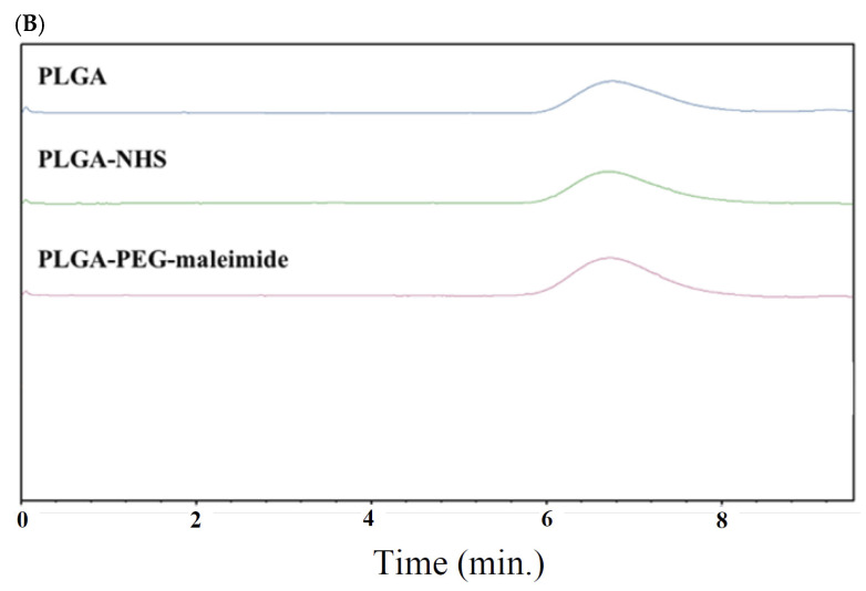
Seliciclib, a cyclin-dependent kinase 5 (CDK5) inhibitor, has demonstrated neuroprotective potential. However, its therapeutic application is limited by poor permeability across the blood-brain barrier (BBB). In this study, polymeric nanoparticles (NPs) modified with a BBB-targeting peptide ligand (His-Ala-Ile-Tyr-Pro-Arg-His) were employed to encapsulate seliciclib. In vitro transport studies showed that the peptide-modified NPs exhibited significantly greater translocation across a bEnd.3 cell monolayer compared to unmodified NPs. Furthermore, in vivo biodistribution analysis revealed that the brain accumulation of peptide-modified NPs was 3.38-fold higher than that of unmodified NPs. Notably, the peptide-conjugated, seliciclib-loaded NPs demonstrated a significant neuroprotective effect against the neurotoxin 1-methyl-4-phenylpyridinium (MPP⁺) in differentiated SH-SY5Y cells.

Keywords: nanoparticles; neuroprotection; peptide; seliciclib.


Conflict of interest statement

The authors declare no conflicts of interest.

Figures

Figure 1
Figure 1
(A) Proton nuclear magnetic resonance (1H-NMR) spectra and (B) size exclusion chromatogram of PLGA-PEG-maleimide.
Figure 1
Figure 1
(A) Proton nuclear magnetic resonance (1H-NMR) spectra and (B) size exclusion chromatogram of PLGA-PEG-maleimide.
Figure 2
Figure 2
(A) Dynamic light scattering (DLS) histograms and (B) TEM images of seliciclib@PPM NPs and seliciclib@PPM NPs-Cys-T7.
Figure 3
Figure 3
Cumulative release of seliciclib from seliciclib@NPs in (A) pH 7.4 phosphate-buffered release medium and (B) pH 5.5 acetate-buffered release medium at 37 °C. (n = 3, mean ± SD).
Figure 4
Figure 4
Transport of free seliciclib, seliciclib@PPM NPs, and seliciclib@PPM NPs-Cys-T7 across bEnd.3 monolayer via an in vitro BBB cell model for 24 h. (n = 3, mean ± SD, *** p < 0.001).
Figure 5
Figure 5
Cellular uptake of peptide-free PP NPs and peptide-conjugated PPM NPs-Cys-T7 in (A) L929 fibroblast cells (as control), (B) bEnd.3 endothelial cells, (C) proliferative SH-SY5Y cells, as well as (D) differentiated SH-SY5Y cells under 5% CO2 at 37 °C for 2 h analyzed by flow cytometry (n = 3, mean ± SD, * p < 0.05, ** p < 0.01, *** p < 0.001, compared to PP NPs), and (E) the increase in cellular uptake of peptide-conjugated NPs relative to peptide-free NPs (calculated by Equation (4)) at 1.5 mg/mL in four cell lines. (F) Flow cytometric analysis of MFI on proliferative and differentiated SH-SY5Y cells stained with anti-human CD71 antibody (green line) and isotype IgG control (orange line), and (G) their CD71 expression levels calculated by Equation (3).
Figure 5
Figure 5
Cellular uptake of peptide-free PP NPs and peptide-conjugated PPM NPs-Cys-T7 in (A) L929 fibroblast cells (as control), (B) bEnd.3 endothelial cells, (C) proliferative SH-SY5Y cells, as well as (D) differentiated SH-SY5Y cells under 5% CO2 at 37 °C for 2 h analyzed by flow cytometry (n = 3, mean ± SD, * p < 0.05, ** p < 0.01, *** p < 0.001, compared to PP NPs), and (E) the increase in cellular uptake of peptide-conjugated NPs relative to peptide-free NPs (calculated by Equation (4)) at 1.5 mg/mL in four cell lines. (F) Flow cytometric analysis of MFI on proliferative and differentiated SH-SY5Y cells stained with anti-human CD71 antibody (green line) and isotype IgG control (orange line), and (G) their CD71 expression levels calculated by Equation (3).
Figure 6
Figure 6
(A) IVIS images of Cy7.5@PPM NPs and Cy7.5@PPM NPs-Cys-T7 in mice brain over 5-day period following intravenous injection via the tail vein. (B) The fluorescence intensity versus time profiles of Cy7.5@NPs accumulated in the brain, and (C) the corresponding area under the curve AUC0–120 h. (D) Ex vivo IVIS images of brains collected 72 h post-injection (n = 3, mean ± SD, * p < 0.05, ** p < 0.01, *** p < 0.001).
Figure 6
Figure 6
(A) IVIS images of Cy7.5@PPM NPs and Cy7.5@PPM NPs-Cys-T7 in mice brain over 5-day period following intravenous injection via the tail vein. (B) The fluorescence intensity versus time profiles of Cy7.5@NPs accumulated in the brain, and (C) the corresponding area under the curve AUC0–120 h. (D) Ex vivo IVIS images of brains collected 72 h post-injection (n = 3, mean ± SD, * p < 0.05, ** p < 0.01, *** p < 0.001).
Figure 7
Figure 7
(A) Cell viability of differentiated SH-SY5Y pretreated with 10 μg/mL of seliciclib, seliciclib@PPM NPs or seliciclib@PPM NPs-Cys-T7 for 2 h, followed by incubation with various concentrations of MPP+ (0.1–2.0 mM) for 48 h, and (B) the corresponding IC50 values were 1.11 ± 0.19 mM, 1.78 ± 0.11 mM and 3.15 ± 0.69 mM, respectively. (n = 3, mean ± SD, ** p < 0.01, *** p < 0.001).

Similar articles

References

    1. Pao P.C., Tsai L.H. Three decades of Cdk5. J. Biomed. Sci. 2021;28:79. doi: 10.1186/s12929-021-00774-y. - DOI - PMC - PubMed
    1. Zhu J., Li W., Mao Z. Cdk5: Mediator of neuronal development, death and the response to DNA damage. Mech. Ageing Dev. 2011;132:389–394. doi: 10.1016/j.mad.2011.04.011. - DOI - PMC - PubMed
    1. Allnutt A.B., Waters A.K., Kesari S., Yenugond V.M. Physiological and pathological roles of CDK5: Potential directions for therapeutic targeting in neurodegenerative diseases. ACS Chem. Neurosci. 2020;11:1218–1230. doi: 10.1021/acschemneuro.0c00096. - DOI - PubMed
    1. Skuntz S., Prochazkova M., Kesavapany S., Amin N.D., Shukla V., Grant P., Kulkarni A.B., Pant H.C. Overex-pression of the Cdk5 inhibitory peptide in motor neurons rescue of amyotrophic lateral sclerosis phenotype in a mouse model. Hum. Mol. Genet. 2019;28:3175–3187. - PMC - PubMed
    1. He F., Qi G., Cai H., Li T., Li M., Zhang Q., Chen J., Ming J., Tian B., Zhang P. Quantitative phosphoproteomic analysis in alpha-synuclein transgenic mice reveals the involvement of aberrant p25/Cdk5 signaling in early-stage Parkinson’s disease. Cell Mol. Neurobiol. 2020;40:897–909. doi: 10.1007/s10571-019-00780-7. - DOI - PMC - PubMed

MeSH terms

LinkOut - more resources