Review
. 2025 Jun 16;26(12):5773.
doi: 10.3390/ijms26125773.

Retinal Autophagy for Sustaining Retinal Integrity as a Proof of Concept for Age-Related Macular Degeneration

Affiliations
Review

Retinal Autophagy for Sustaining Retinal Integrity as a Proof of Concept for Age-Related Macular Degeneration

Roberto Pinelli et al. Int J Mol Sci. .

Abstract

Current evidence indicates that most types of autophagy represent a pivot in promoting retinal integrity. In healthy conditions, autophagy acts on multiple pathways, which are fundamental for the biochemistry and the fine structure of the retina. Autophagy is essential in granting visual processes. On the other hand, autophagy dysfunction characterizes several retinal disorders. This is mostly evident in age-related macular degeneration (AMD), which represents the most common degenerative disease leading to blindness. The involvement of autophagy in AMD is documented in vitro and in vivo experiments, and it is strongly suggested by clinical findings in humans. The present manuscript provides an overview of the specific types of autophagy, which prevail in the retina and their alterations in retinal degeneration with an emphasis on AMD. The dysfunction of specific autophagy steps was analyzed in relation to hallmarks of AMD pathology and symptoms. An extended session of the manuscript analyzes the connection between altered autophagy and cell pathology within retinal pigment epithelium, as well as the site and structure of extracellular aggregates named drusen. The significance of the drusen in relation to visual function is discussed in the light of the role of autophagy in regulating key steps of phototransduction.

Keywords: autophagoproteasome; drusen; lipophagy; lysosome; mitophagy; proteasome; pseudodrusen; retinal degeneration; retinal neurovascular unit; retinal pigment epithelium.


Conflict of interest statement

The authors declare no conflicts of interest.

Figures

Figure 1
Figure 1
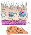
Introductory graphical scheme of various types of autophagy and various retinal disorders. The concept of autophagy as a relevant pathway in the retina is rather generic since multiple types of autophagy exist, and their role may vary depending on the specific retinal layer and disorders. The cartoon reporting the prototype of autophagy vacuoles is also drawn according to a general scheme, which includes the removal of various chemical species (proteins, lipids, carbohydrates, nucleic acids) and organelles (mitochondria, endoplasmic reticulum). Within specific retinal disorders, which are featured by altered autophagy, a difference may exist concerning specific types of autophagy. It is likely that the occurrence of retinal damage following various pathological conditions of the eye may be produced by a defect in distinct autophagy steps. The effort to distinguish between various forms of autophagy and to dissect which kind is most relevant in AMD represents a major aim of the present manuscript.
Figure 2
Figure 2
Autophagy operates within a healthy retina, and its alteration occurs at late stages of most retinal degenerative disorders. Retinal degenerative disorders featuring early loss of photoreceptors consist of several forms of retinal degenerative diseases. In these disorders, which start at the level of the outer retina, where RPE and the outer segment of the photoreceptor are placed, a progression downstream to the inner retina often occurs according to a common convergent pathology. This recruits various cell layers of the retinal circuitry to reach the ganglionic cell layer, as indicated in the figure labelling. This produces retinal maladaptive plasticity [23], where alterations of retinal structure are bound to autophagy failure [9,16,23]. At the final stage, the widespread retinal pathology mimics central neurodegenerative disorders [23,24,25]. ELM = external limiting membrane; ILM = inner limiting membrane; PIS = photoreceptor inner segment; POS = photoreceptor outer segment.
Figure 3
Figure 3
Scheme of RPE autophagy reporting sites where autophagy dysfunction triggers cell pathology in AMD. The quite generic pattern of physiological retinal autophagy progression consists of the shuttling of the lipid membrane of the photoreceptor outer segment (POS), along with other lipid content within lipid droplets, entering the nascent autophagosome (phagophore), which is stained for the presence of microtubule-associated light chain protein 3 (LC3). Within autophagosomes lipids are stored along with misfolded proteins, glycogen metabolic by-products, altered mitochondria and other constituents mentioned in the text. The relevant entry of lipid droplets and POS may occur directly into lysosomes, which otherwise merge with autophagosomes. The clearance of these constituents in the lysosome may be defective in the course of AMD. This generates abundant lipid droplets and glycogen granules along with culprit proteins within RPE cells. Again, this defective clearance leads to activate a non-canonical autophagy step, which leads to autophagy-dependent exocytosis, where lysosomes, multivesicular bodies (MVBs) or other membrane limited organelles are extruded either towards the Bruch’s membrane (likely generating drusen) or towards the opposite domain, which intermingles with the outer segment of photoreceptors (likely generating pseudodrusen).
Figure 4
Figure 4
The generation of drusen and pseudodrusen in AMD. Site- and structure-specificity of drusen is closely related to RPE [48]. Drusen are produced mainly by secretory autophagy, where cell cargoes are released via the plasma membrane [47]. Drusen accumulate within the thin space underlying the basal membrane of RPE, just above the Bruch’s membrane, or alternatively, the space between RPE and the outer segment of the photoreceptors (pseudodrusen). The structure of drusen and pseudodrusen matches the content of all lysosomes and MVBs.
Figure 5
Figure 5
Imaging of the right retina as a representative image of AMD. (A) The occurrence of drusen, below the RPE between retina and choroid, is visualized in a representative optical coherence tomography (OCT) from a 68-year-old woman, who gave her informed consent. In addition, from the same patient, the retinal cube is presented (B). This corresponds to a computer-aided manipulation of the macular cube, which is defined by the volume of the macular region that equals 1 mm3. In the macular cube, it is possible to count drusen based on a single plane obtained by OCT, which transforms the region of interest from a volume into an area. The retinal pigment epithelium is evidenced in red by computerized processing in the macular cube. The occurrence of drusen in the OCT is expressed by red arrows. At this level, it is also evident that the thinning of the RPE and the decreased length of the photoreceptor layer (indicated by red asterisks) above the drusen. The patient suffers from a loss of visual acuity (best corrected visual acuity, BCVA = 20/50) and she perceives spread longitudinal temporal metamorphopsia.
Figure 6
Figure 6
The role of retinal autophagy in removing altered proteins. The natural progression of protein autophagy consists of the binding of an altered/misfolded protein structure with ubiquitin protein or a chain of poly-ubiquitin in the process named protein ubiquitination [54]. The latter process received specific attention recently, since protein ubiquitination is required to address protein clearance both via the proteasome and autophagy pathways. In detail, the recruitment of altered proteins by the proteasome is based on the poly-ubiquitination, while poly/mono-ubiquitination addresses altered proteins to the autophagy vacuoles. As recently shown, and described in further detail in the main text, the proteasome itself, bound to a poly-ubiquitin chain and a misfolded protein, is fully shuttled within the autophagosome. This occurs massively following autophagy activation (via mTOR inhibition) to form a merged clearing organelle named autophagoproteasome through a shuttle protein named p62 (sequestosome), which binds the vesicle membrane, mainly through its Uba domain. As a result, an empowerment of enzyme activity is obtained by adding proteasome proteolytic enzymes to the enzymes occurring within the lysosomal compartment, indicated by the lysosome-associated membrane protein 1 (LAMP1).
Figure 7
Figure 7
Specificity of retinal lipid autophagy (lipophagy). In the retina, lipophagy consists of a specific activity of the autophagy machinery leading to degradation of the photoreceptor outer segments (POS), which contribute largely to forming lipid droplets within RPE cells. The conspicuous amount of lipid droplets is taken up by the lysosomal compartment upon the merging of lysosomal vacuoles and lipid vacuoles/droplets. Very often, the lipid droplets directly enter the lysosomes and MVBs, although the specific receptors remain questionable. Some recent evidence suggests that the protein family forming the vacuolar protein sorting required for transfer (VPS), such as VPS4A found in the liver, may be strongly involved, along with the early autophagy inducer beclin1. One might consider that in baseline conditions, retinal autophagy, while granting the clearance of some misfolded proteins, is strongly engaged in taking up and degrading innumerable lipid droplets. When autophagy is impeded some protein aggregates persist although the lipids engulf the cell. This explains why the secretory autophagy of lipids leads to the formation of drusen, where lipids represent almost half of the whole volume.
Figure 8
Figure 8
Representative staining of lipid droplets following autophagy inhibition (3-methyladenine, 3-MA, 10 mM) in human RPE cell line (ARPE). The staining of lipids was obtained with Sudan Black B (8 min exposure), while nuclei were counterstained with a Nissl-type approach (Fast Red, 7 min). Scale Bar = 15 μm. Original picture by M.F. and G.L.
Figure 9
Figure 9
Lipophagy within RPE cells. Lipid droplets are well evident within RPE cells in baseline conditions and are in excess during AMD. Lipid autophagy is fundamental in maintaining the viability of RPE cells since lipids are abundant in the physiology of RPE. Several systemic sources, such as the bloodstream and lymphatic stream in the choroid of patients affected by dyslipidemia, deliver lipids to the RPE. However, the greatest amount of lipids is specifically driven by an excess of lipid substrates entering the RPE from the outer segment of photoreceptors (POS). A major constituent of photosensitive disks is retinoic acid in the form of its aldehyde. Remarkably, all-trans aldehyde derived from retinoic acid (all-trans-retinoic acid, ATRA) stimulates autophagy. These lipids may enter either the autophagosome or the lysosomal compartment. Indeed, when lipids are in excess and coalesce to form lipid droplets, the preferential pathway consists of the direct entry into lysosomes, where the VPS-related receptor is the candidate receptor for lipid droplets. The storage of lipid-rich lysosomes, lipid-rich VPS, and frankly abundant interspersed lipid droplets within RPE cells is likely to trigger non-conventional secretion and non-canonical secretory autophagy to build up at least half of the whole drusen volume.
Figure 10
Figure 10
Dual effects of lipids on retinal autophagy. While lipids are substantial substrates for the autophagy within RPE cells, which mostly recruits a direct lysosome clearance, the activity of some phospholipids, such as phophatidylethanolamine (PEA), is a powerful inducer of LAP autophagy. PEA generates the lipidation of LC3, which feeds the nascent phagophore membrane and modulates its membrane bending, leading to ceiled autophagosomes.
Figure 11
Figure 11
The sphingolipid ceramide stimulates autophagy by acting on Beclin1. Ceramide promotes the removal of Beclin1 from the Beclin1/Bcl-2 complex. This finally leads to the formation of a multi-protein complex (known as Beclin1-VPS34-VPS15), which stimulates autophagy [102].
Figure 12
Figure 12
Glycogen-specific autophagy (glycophagy). Glycogen chains in the cell are built up by adding moieties of uridine diphosphate (UDP)-glucose as a substrate. These primary chains of glucose become very long through the progressive addition of subunits through the enzymatic activity of glycogen synthase. The main chain is further branched in its secondary chains by the activity of glycogen-branching enzyme. The autophagy degradation of these glycogen long chains occurs via recognition with a specific receptor for glycogen, named starch binding domain-containing protein 1 (STBD1). This binding addresses cytosolic glycogen towards the autophagy machinery (glicophagy), once it is stored within LC3 and beclin1-positive autophagosomes. In this way, a specific phagosome is formed, which is named glycophagosome. Following autophagosome–lysosome fusion, a specific enzyme named α-glucosidase mediates lysosomal glycogen breakdown [112,113]. Single glucose molecules may enter directly into the lysosome through a specific glucose-sensing site on the lysosome.

Similar articles

References

    1. Markitantova Y., Simirskii V. Retinal Pigment Epithelium Under Oxidative Stress: Chaperoning Autophagy and Beyond. Int. J. Mol. Sci. 2025;26:1193. doi: 10.3390/ijms26031193. - DOI - PMC - PubMed
    1. Bergmann M., Schütt F., Holz F.G., Kopitz J. Inhibition of the ATP-driven proton pump in RPE lysosomes by the major lipofuscin fluorophore A2-E may contribute to the pathogenesis of age-related macular degeneration. FASEB J. 2004;18:562–564. doi: 10.1096/fj.03-0289fje. - DOI - PubMed
    1. Kaushal S. Effect of rapamycin on the fate of P23H opsin associated with retinitis pigmentosa (an American Ophthalmological Society thesis) Trans. Am. Ophthalmol. Soc. 2006;104:517–529. - PMC - PubMed
    1. Kunchithapautham K., Rohrer B. Apoptosis and autophagy in photoreceptors exposed to oxidative stress. Autophagy. 2007;3:433–441. doi: 10.4161/auto.4294. - DOI - PubMed
    1. Mellén M.A., de la Rosa E.J., Boya P. The autophagic machinery is necessary for removal of cell corpses from the developing retinal neuroepithelium. Cell Death Differ. 2008;15:1279–1290. doi: 10.1038/cdd.2008.40. - DOI - PubMed

LinkOut - more resources