. 2025 Jun 16;26(12):5775.
doi: 10.3390/ijms26125775.

Biotype Determines Survival of Yersinia enterocolitica in Red Blood Cell Concentrates

Affiliations

Biotype Determines Survival of Yersinia enterocolitica in Red Blood Cell Concentrates

Katarzyna Morka et al. Int J Mol Sci. .

Abstract

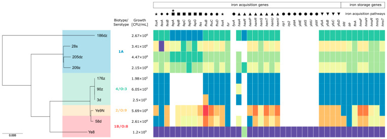
Red blood cell (RBC) concentrates remain at risk of bacterial contamination during cold storage. Although infrequent, Yersinia enterocolitica poses a significant blood safety risk. This study aimed to assess Y. enterocolitica bioserotype growth in RBC concentrates, serum sensitivity, and genetic diversity including iron metabolism genes. Ten Y. enterocolitica isolates from bioserotypes 1A, 1B/O:8, 4/O:3, and 2/O:9 were incubated in RBC concentrates and counted on days 3, 7, 14, 21, and 28. After incubation, the isolates were tested in human serum (NHS). Eight genomes were sequenced, analyzed using cgMLST, and screened for iron metabolism genes. The isolates formed two clusters, with 186dz (1A) and Ye8 (1B/O:8) as singletons. After 28 days in the RBC concentrates, the bacterial counts ranged from 1.98 × 10⁵ to 1.2 × 10⁹ CFU/mL, with Ye8 (1B/O:8) achieving the highest growth and one 4/O:3 isolate showing the lowest. All isolates survived 15-30 min in NHS, but the 28s isolate did not survive at 60 min. Serum sensitivity increased in two isolates, decreased in three, and remained unchanged in five. Isolates contained 27-42 iron metabolism genes with multiple allelic variants. The iron metabolism gene content or variants may influence the growth of Y. enterocolitica in RBC.

Keywords: Yersinia enterocolitica; bioserotypes; blood safety; contamination; red blood cells.


Conflict of interest statement

The authors declare no conflicts of interest.

Figures

Figure 1
Figure 1
Phylogenetic tree of the studied Y. enterocolitica isolates, alongside the distribution of 42 iron metabolism-related genes and their growth potential in red blood cells (RBCs) after 28 days of incubation. The tree was constructed using the core genome multilocus sequence typing (cgMLST) scheme and the Jukes and Cantor evolutionary model, visualized with Phandango. Each isolate is annotated with its corresponding iron metabolism-related gene repertoire. Genes are represented by colored blocks indicating different allelic variants, while white blocks signify gene absence. Clades in the phylogenetic tree highlight genetic relationships among isolates including differences in bioserotypes, allelic repertoires, and the presence or absence of specific genes. The number of detected genes in each isolate ranged from 27 to 42, reflecting variation in their iron acquisition capabilities. Reference strains Ye8 and Ye9N are included for comparison, with Ye8 harboring all 42 genes. Clustering patterns correlate with specific genetic characteristics. Legend:●—yersiniabactin (Ybt), ∎—enterochelin catecholate siderophore, ▲—Hem uptake system, ★—TonB-dependent OM transporters, ◆—Feo system, ▼—inorganic iron ABC transport systems of Y. pestis (Yfu), ⬟—ferric hydroxamate uptake system (fhuCDB), fur—the master regulator of iron homeostasis in Y. enterocolitica, bfr—bacterioferritin, bfd—bacterioferritin-associated ferredoxin complex, ftn—ferritin, hmsFRST—hemin storage system.
Figure 2
Figure 2
Bacterial growth in triplicates in RBC concentrates during 28 days of incubation at 2–4 °C. The data from each RBC incubation were compared by analysis of variance (two-way ANOVA). Bacterial survival rates were compared using the Tukey’s multiple comparisons test. The p-values from these tests are shown in the text. p < 0.05 indicates that the compared values were significantly different at a 95 % confidence level.
Figure 3
Figure 3
Survival of pathogenic and nonpathogenic 1A Y. enterocolitica isolates during 60 min at 37 °C in NHS before and after RBC concentrate incubation. The asterisks indicate statistically significant differences between strains (ns p > 0.05, ** p < 0.01, *** p < 0.001, **** p < 0.0001, by two-way ANOVA with Tukey’s comparison post-test).

Similar articles

References

    1. Guinet F., Carniel E., Leclercq A. Transfusion-transmitted Yersinia Enterocolitica sepsis. Clin. Infect. Dis. 2011;53:583–591. doi: 10.1093/cid/cir452. - DOI - PubMed
    1. Prax M., Spindler-Raffel E., McDonald C.P., Bearne J., Satake M., Kozakai M., Rojo J., Hanschmann K.O., Lambrecht B., Grundmann U., et al. Establishment of Transfusion-relevant Bacteria Reference Strains for Red Blood Cells. Vox Sang. 2021;116:692–701. doi: 10.1111/vox.13057. - DOI - PubMed
    1. Shepel M., Boyd J., Luider J., Gibb A.P. Interaction of Yersinia enterocolitica and Y. Pseudotuberculosis with Platelets. J. Med. Microbiol. 2001;50:1030–1038. doi: 10.1099/0022-1317-50-12-1030. - DOI - PubMed
    1. Ramirez-Arcos S., Kou Y., Cayer M., De Grandmont M., Girard M., Cloutier M. The Impact of Red Blood Cell Manufacturing Variables on Bacterial Growth Dynamics: A Pilot Study. Vox Sang. 2019;114:478–486. doi: 10.1111/vox.12782. - DOI - PubMed
    1. Hoelen D.W.M., Tjan D.H.T., Schouten M.A., Dujardin B.C.G., van Zanten A.R.H. Severe Yersinia Enterocolitica Sepsis after Blood Transfusion. Neth. J. Med. 2007;65:301–303. - PubMed

MeSH terms

LinkOut - more resources