. 2025 Jun 17;26(12):5799.
doi: 10.3390/ijms26125799.

Kringle-Dependent Inhibition of Plasmin-Mediated Fibrinolysis by Native and Citrullinated Core Histones

Affiliations

Kringle-Dependent Inhibition of Plasmin-Mediated Fibrinolysis by Native and Citrullinated Core Histones

Erzsébet Komorowicz et al. Int J Mol Sci. .

Abstract

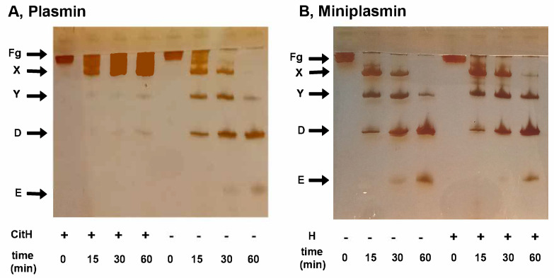
The fibrin matrix of thrombi is intertwined with neutrophil extracellular traps (NETs) containing histones that render resistance to fibrinolysis. During NET formation, histones are citrullinated. Our study addresses the question of whether citrullination modifies the fibrin-stabilizing effects of histones. We studied the structure and viscoelastic properties of fibrin formed in the presence of native or citrullinated H1 and core histones by scanning electron microscopy, clot permeation, and oscillation rheometry. The kinetics of fibrin formation and its dissolution were followed by turbidimetry and thromboelastometry. Co-polymerizing H1 with fibrin enhanced the mechanical strength of the clots, thickened the fibrin fibers, and enlarged the gel pores. In contrast, the addition of core histones resulted in a reduction in the fiber diameter, and the pores were only slightly larger, whereas the mechanical stability was not modified. Plasmin-mediated fibrinogen degradation was delayed by native and citrullinated core histones, but not by H1, and the action of des-kringle1-4-plasmin was not affected. Plasmin-mediated fibrinolysis was inhibited by native and citrullinated core histones, and this effect was moderated when the kringle domains of plasmin were blocked or deleted. These findings suggest that in NET-containing thrombi that are rich in core histones, alternative fibrinolytic enzymes lacking kringle domains are more efficient lytic agents than the classic plasmin-dependent fibrinolysis.

Keywords: fibrin; neutrophil extracellular traps; thrombosis.


Conflict of interest statement

The authors declare no conflicts of interest.

Figures

Figure 1
Figure 1
Mechanical stabilization of fibrin structure by H1, as opposed to core histones. H1 (red) or core (blue) histones at 25 mg/L were added to 7.4 µM fibrinogen and the mixture was clotted with 8 nM thrombin for 12 min under oscillatory shear in the measurement gap of the rheometer, as detailed in Materials and Methods. Thereafter, the clots were subjected to stepwise increasing shear stress, while the resulting strain was continuously monitored. Panel (A) shows the dynamic viscosity values calculated as the ratio of stress/strain. Panel (B) shows the relative deformation. Black lines depict the viscoelastic behavior of pure fibrin gel for comparison. The presented curves are representatives of a series of experiments (n = 4–8); numerical data and statistics are shown in Table 2.
Figure 2
Figure 2
Plasmin-mediated fibrinolysis of composite fibrin/histone clots in extrinsic and intrinsic models. (A): Extrinsic lysis model: fibrinogen (6 µM) was clotted with 2 nM thrombin in the absence (black) or presence of 100 mg/L native (solid line) or citrullinated (dashed line) H1 (red) or core (blue) histones, and after a 2 h clotting period, 500 nM plasmin was layered on the clot surface to initiate fibrin dissolution. (B): Intrinsic lysis model: fibrinogen (6 µM) containing none (black) or 75 mg/L native (solid line) or citrullinated (dashed line) H1 (red) or core (blue) histones was clotted with 10 nM thrombin with 8 nM plasmin added at the same time to induce fibrinolysis simultaneously with clotting. The acending and descending parts of the turbidimetric curve recorded as absorbance at 340 nm wavelength indicate fibrin formation and dissolution. The presented kinetic curves are averages from 5 parallel measurements on the same day and were normalized considering maximal turbidity as 1.
Figure 3
Figure 3
Histones inhibit plasmin-mediated fibrinolysis in a kringle-dependent manner. Fibrinogen (6 µM) containing native (solid line) or citrullinated (dashed line) H1 (red) or H3 (blue) histones at indicated concentrations was clotted with 10 nM thrombin in the presence of 8 nM plasmin (panel (A)), kringle-blocked plasmin (panel (B)) or miniplasmin (panel (C)), or 26 nM elastase (panel (D)) added at the same time to induce fibrinolysis simultaneously with clotting. Lysis time (LT50) was computed on the descending part of the recorded turbidimetric curve in a similar setup, as illustrated in Figure 2B, and plotted in relative units (RU) considering LT50 in the absence of histones as a reference (43 min, 78 min, 98 min and 37 min for plasmin, kringle-blocked plasmin, miniplasmin, and elastase, respectively). Data are plotted as mean ± SD values (n = 10–15).
Figure 4
Figure 4
Plasmin- and miniplasmin-mediated loss of fibrinogen clottability in the presence of native or citrullinated histones. Fibrinogen (6 µM) was incubated with 12 nM plasmin (A) or miniplasmin (B), and thrombin at concentration set to yield a 10 s clotting time with undegraded fibrinogen was added to samples withdrawn after various times to assess the presence of residual clottable fibrinogen. Samples with clotting times longer than 120 s were plotted as 121 s and considered non-clottable due to extensive fibrinogen degradation. Additives in the panels are as follows: (A) Native (solid) or citrullinated (dashed) H1 (red) or core (blue) histones at 50 mg/L (B): Native H1 (red) or core (blue) histones at 50 mg/L concentrations; control curves are all black. Representative curves are presented, which were repeated on at least 3 different days to verify the observed tendencies.
Figure 5
Figure 5
Formation of fibrinogen degradation products by plasmin or miniplasmin in the presence of histones. Fibrinogen (1.5 µM) was incubated with 12 nM plasmin (panel (A)) or miniplasmin (panel (B)) in the absence or presence of 50 mg/L native (B) or citrullinated (A) core histones. At time intervals, the samples were withdrawn, heated and subjected to SDS-PAGE in a 7.5% homogeneous gel, followed by silver staining. Band labels: Fg, fibrinogen; X/Y/D/E, fibrinogen degradation products.

Similar articles

References

    1. Khorasanizadeh S. The nucleosome. From genomic organization to genomic regulation. Cell. 2004;116:259–272. doi: 10.1016/S0092-8674(04)00044-3. - DOI - PubMed
    1. Millán-Zambrano G., Burton A., Bannister A.J., Schneider R. Histone post-translational modifications—Cause and consequence of genome function. Nat. Rev. Genet. 2022;23:563–580. doi: 10.1038/s41576-022-00468-7. - DOI - PubMed
    1. Andrés M., Garcia-Gomis D., Ponte I., Suau P., Roque A. Histone H1 Post-Translational Modifications: Update and Future Perspectives. Int. J. Mol. Sci. 2020;21:5941. doi: 10.3390/ijms21165941. - DOI - PMC - PubMed
    1. Arita K., Shimizu T., Hashimoto H., Hidaka Y., Yamada M., Sato M. Structural basis for histone N-terminal recognition by human peptidylarginine deiminase 4. Proc. Natl. Acad. Sci. USA. 2006;103:5291–5296. doi: 10.1073/pnas.0509639103. - DOI - PMC - PubMed
    1. Hagiwara T., Hidaka Y., Yamada M. Deimination of histone H2A and H4 at arginine 3 in HL-60 granulocytes. Biochemistry. 2005;44:5827–5834. doi: 10.1021/bi047505c. - DOI - PubMed

LinkOut - more resources