. 2025 Jun 18;26(12):5857.
doi: 10.3390/ijms26125857.

Distinctive Features of Extracellular Vesicles Present in the Gastric Juice of Patients with Gastric Cancer and Healthy Subjects

Affiliations

Distinctive Features of Extracellular Vesicles Present in the Gastric Juice of Patients with Gastric Cancer and Healthy Subjects

Gleb Skryabin et al. Int J Mol Sci. .

Abstract

Extracellular vesicles (EVs) are key mediators of intercellular communication and play a vital role in cancer progression. While EVs in the blood are well-studied, those in local body fluids, such as gastric juice (GJ), remain underinvestigated. Previously, we first characterized GJ-derived EVs and demonstrated their potential for gastric cancer (GC) screening. Here, we conducted a detailed morphological analysis of GJ-EVs using cryo-electron microscopy, identifying both typical and atypical EV subtypes, and categorized their relative abundances. A subsequent comparison of the size distribution of GJ-derived EVs by nanoparticle tracking analysis revealed significant differences between samples obtained from GC patients (n = 40) and healthy subjects (n = 25). Additionally, the mean EV sizes differed significantly according to the presence of the tetraspanin protein CD9. Furthermore, the ratio of CD9-positive to CD9-negative EV samples differed between cancer patients and healthy donors. These data suggest that GJ contains distinct subpopulations of EVs that vary in size and CD9 expression, as well as EVs with certain types of atypical morphology. The identification of discrepancies in EV size and the presence of CD9 between GJ from cancer patients and healthy individuals offers potential avenues for the identification of new GC markers.

Keywords: CD9; cryo-electron microscopy; exosomes; gastric cancer; gastric juice; nanoparticle tracking analysis; small extracellular vesicles; tetraspanins.


Conflict of interest statement

The authors declare no conflicts of interest.

Figures

Figure 1
Figure 1
Visualization of GJ EVs by TEM. (A) TEM images depicting particles with typical EV size and morphology from GC patients and healthy donors. (B) Examples of vesicles with atypical morphology visualized by TEM.
Figure 2
Figure 2
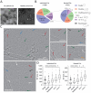
(A) Diversity of morphological types of GJ EVs. (B) The prevalence of each EV morphological subcategory, n = sample size. (C) Cryo-EM images illustrating the morphological diversity of GJ EVs. Micrographs of single (white arrows), double (red), multilayered (green), “2-in-1” (blue), “>2-in-1” (black), and dumbbell-shaped (orange) vesicles are shown. Scale bars 200 nm. (D) Sizes of each individual (left) and nested (right) EV morphological subcategory (**** p < 0.0001, * p < 0.05, ns—not significant). Graphs are plotted using the Tukey method.
Figure 3
Figure 3
EV size distribution. (A) The size distribution histograms of the distinct EV morphology subtypes of ‘individual’ EVs (top row) and ‘nested’ EVs (bottom row). Data were obtained from cryo-EM images. n = sample size. (B) Comparison of size distribution data obtained from NTA (left) and cryo-EM (right). For distribution based on cryo-EM, only ‘individual’ EVs were included in the graph. Symbol ± indicates SEM.
Figure 4
Figure 4
Characterization of EVs isolated from gastric juice. (A) Western blot analysis of exosomal markers flotillin-2, TSG101, stomatin, and CD9 in GC and HD EVs. Tumor cell lysate (TCL) obtained from gastric adenocarcinoma SNU-1 cell line was used as a control. The PCNA protein was used to confirm the absence of cellular proteins of non-vesicular origin in EV preparations. (B) NTA data on mean EV size distribution of a population of CD9(+) and CD9(–) samples. Error bars indicate SEM. (C) Mean values for EV size characteristics of the CD9(+) and CD9(–) samples and entire data according to NTA data. (D) Comparison of the mean size of CD9(+) and CD9(–) EVs (** p < 0.01).
Figure 5
Figure 5
The characterization of GJ-derived EVs by nanoparticle tracking analysis. (A) NTA data on the mean EV size distribution of a population of gastric cancer (GC) and healthy donor (HD) samples. Error bars indicate SEM. (B) Mean values for EV size characteristics over the GC and HD samples according to NTA data. (C) Comparison of the mean size of GC and HD EVs. Bars indicate min to max. (D) Distribution of CD9(+) and CD9(–) EVs over the GC and HD groups. (E) Comparison of the mean size of CD9(+) and CD9(–) EVs in the GC and HD groups. The graph is plotted using the Tukey method.

Similar articles

References

    1. Bray F., Laversanne M., Sung H., Ferlay J., Siegel R.L., Soerjomataram I., Jemal A. Global Cancer Statistics 2022: GLOBOCAN Estimates of Incidence and Mortality Worldwide for 36 Cancers in 185 Countries. CA Cancer J. Clin. 2024;74:229–263. doi: 10.3322/caac.21834. - DOI - PubMed
    1. Correa P. Gastric Cancer: Overview. Gastroenterol. Clin. N. Am. 2013;42:211–217. doi: 10.1016/j.gtc.2013.01.002. - DOI - PMC - PubMed
    1. Li Y., Feng A., Zheng S., Chen C., Lyu J. Recent Estimates and Predictions of 5-Year Survival in Patients with Gastric Cancer: A Model-Based Period Analysis. Cancer Control. 2022;29:107327482210992. doi: 10.1177/10732748221099227. - DOI - PMC - PubMed
    1. Sekiguchi M., Oda I., Matsuda T., Saito Y. Epidemiological Trends and Future Perspectives of Gastric Cancer in Eastern Asia. Digestion. 2021;103:22–28. doi: 10.1159/000518483. - DOI - PubMed
    1. Li Y., He X., Li Q., Lai H., Zhang H., Hu Z., Li Y., Huang S. EV-Origin: Enumerating the Tissue-Cellular Origin of Circulating Extracellular Vesicles Using exLR Profile. Comput. Struct. Biotechnol. J. 2020;18:2851–2859. doi: 10.1016/j.csbj.2020.10.002. - DOI - PMC - PubMed

LinkOut - more resources