. 2025 Jun 19;26(12):5865.
doi: 10.3390/ijms26125865.

Long Non-Coding RNA LOC401312 Induces Radiosensitivity Through Upregulation of CPS1 in Non-Small Cell Lung Cancer

Affiliations

Long Non-Coding RNA LOC401312 Induces Radiosensitivity Through Upregulation of CPS1 in Non-Small Cell Lung Cancer

Zhengyue Cao et al. Int J Mol Sci. .

Abstract

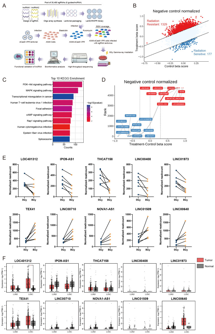
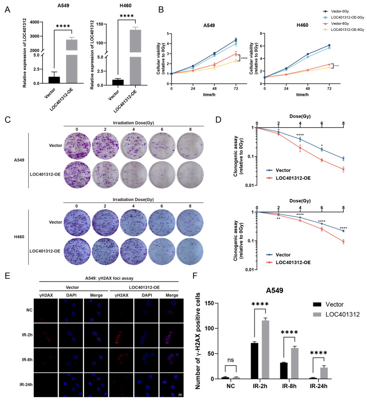
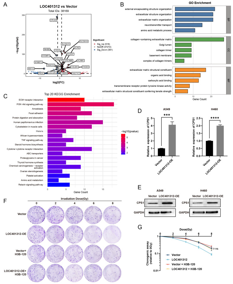
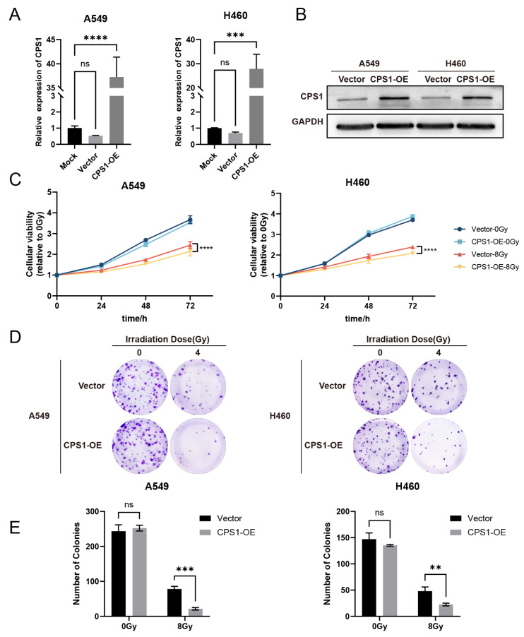
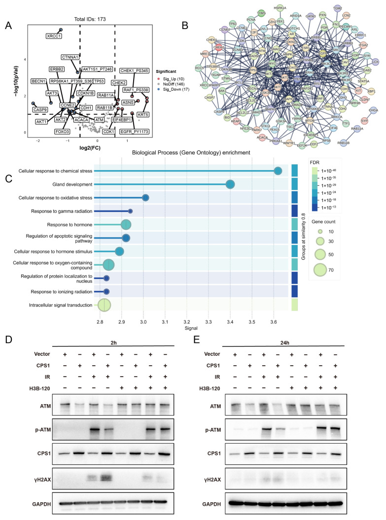
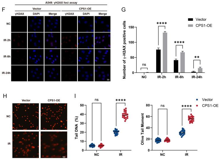
Long noncoding RNAs (lncRNAs), non-protein-coding transcripts exceeding 200 nucleotides, are critical regulators of gene expression through chromatin remodeling, transcriptional modulation, and post-transcriptional modifications. While ionizing radiation (IR) induces cellular damage through direct DNA breaks, reactive oxygen species (ROS)-mediated oxidative stress, and bystander effects, the functional involvement of lncRNAs in the radiation response remains incompletely characterized. Here, through genome-wide CRISPR activation (CRISPRa) screening in non-small cell lung cancer (NSCLC) cells, we identified LOC401312 as a novel radiosensitizing lncRNA, the stable overexpression of which significantly enhanced IR sensitivity. Transcriptomic profiling revealed that LOC401312 transcriptionally upregulates carbamoyl-phosphate synthase 1 (CPS1), a mitochondrial enzyme involved in pyrimidine biosynthesis. Notably, CPS1 overexpression recapitulated the radiosensitization phenotype observed with LOC401312 activation. Mechanistic investigations revealed that CPS1 suppresses the phosphorylation of ATM kinase (Ser1981) protein, which is a key mediator of DNA damage checkpoint activation. This study established the LOC401312-CPS1-ATM axis as a previously unrecognized regulatory network governing radiation sensitivity, highlighting the potential of lncRNA-directed metabolic rewiring to impair DNA repair fidelity. Our findings not only expand the functional landscape of lncRNAs in DNA damage response but also provide a therapeutic rationale for targeting the LOC401312-CPS1 axis to improve radiotherapy efficacy in NSCLC.

Keywords: CPS1; CRISPRa screening; LOC401312; ionizing radiation sensitivity regulation; long noncoding RNA.


Conflict of interest statement

The authors declare no conflicts of interest.

Figures

Figure 1
Figure 1
Systematic Identification of Radiation-Responsive lncRNAs via CRISPRa Screening in NSCLC. (A) Schematic of the CRISPRa high-throughput library screening strategy for identifying radiation-responsive lncRNAs in A549 cells, comprising 30,480 sgRNAs targeting 4766 lncRNAs (6 sgRNAs per gene) with >500× coverage per sgRNA. (B) Scatter plot of Delta-beta scores for screened lncRNAs. (C) KEGG pathway analysis and protein interaction network of candidate lncRNAs predicted using the LncSEA database (http://bio.liclab.net/LncSEA, accessed on 1 November 2021). (D) Delta-beta score ranking and differential gene visualization of identified lncRNAs. (E) Enrichment analysis of sgRNAs targeting lncRNAs, showing significant depletion of all six sgRNAs (normalized read counts shown). (F) Differential lncRNAs expression in normal tissues versus lung adenocarcinoma (LUAD) and squamous cell carcinoma (LUSC) clinical samples using the GEPIA2 database (http://gepia2.cancer-pku.cn, accessed on 10 June 2025). * p < 0.05 by unpaired two-tailed Student’s t-test.
Figure 2
Figure 2
LOC401312 Validated as a Radiosensitizing lncRNA in Lung Cancer Cells. (A) LOC401312 overexpression validation. The RT-qPCR results were normalized against GAPDH expression. Data are presented as mean ± SD of three biologically independent replicates. **** p < 0.0001 by unpaired two-tailed Student’s t-test. (B) CCK-8 cellular viability curves of LOC401312-overexpressing cells post 8 Gy irradiation. Data are presented as mean ± SD of five biologically independent replicates. **** p < 0.0001 by two-way ANOVA. (C) Clonogenic survival assays at 0, 2, 4, 6, and 8 Gy (14-day post-irradiation, n = 3 biological replicates, the diameter of each individual image is 35 mm). (D) Quantification of clonogenic survival rates. Data are presented as mean ± SD * p < 0.05, ** p < 0.01, **** p < 0.0001 by two-way ANOVA. (E) Immunofluorescence imaging of γH2AX foci in LOC401312-overexpressing cells at 2 h, 8 h and 24 h post 8 Gy irradiation. Representative images of three independent experiments (n = 3) are shown (blue—DAPI, red—γH2AX. Scale bar: white, 10 μm). (F) Statistical analysis of number of γH2AX positive foci per cell. **** p < 0.0001 by two-way ANOVA. ns, not significant.
Figure 3
Figure 3
Transcriptomic Profiling of LOC401312-Overexpressing A549 Cells Identifies CPS1 as a Functional Mediator of Radiation Sensitivity. (A) Volcano plots show the -log normalized p-value and log2 fold change of transcriptomic profiling in LOC401312-overexpressing A549 cells compared to the vector control. The upregulated and downregulated DEGs are red and blue, respectively, and the undifferentiated genes expressed in both groups are denoted in gray. (B,C) GO biological process (B) and KEGG (C) analysis of DEGs in (A). (D) qRT-PCR validation of CPS1 mRNA upregulation. The RT-qPCR results were normalized against GAPDH expression. Data are presented as mean ± SD of three biologically independent replicates. *** p < 0.001, **** p < 0.0001 by unpaired two-tailed Student’s t-test. (E) Western blot confirmation of CPS1 protein induction. (F) Clonogenic survival assays of LOC401312-overexpressing cells irradiated with 0–8 Gy, with 10 μM H3B-120 (CPS1 inhibitor) pretreatment for 48 h (14-day post-irradiation, n = 3 biological replicates, the diameter of each individual image is 35 mm). (G) Survival fraction quantification showing abolished radiation sensitization by H3B-120; ns, not significant.
Figure 4
Figure 4
CPS1 overexpression induces radiation sensitivity in lung cancer cells. (A) qRT-PCR validation of CPS1 mRNA upregulation. The RT-qPCR results were normalized against GAPDH expression. Data are presented as mean ± SD of three biologically independent replicates. *** p < 0.001, **** p < 0.0001 by one-way ANOVA. (B) Western blot analysis of CPS1 protein levels. (C) CCK-8 cellular viability curves of CPS1-overexpressing cells post 8 Gy irradiation. Data are presented as mean ± SD of five biologically independent replicates. **** p < 0.0001 by two-way ANOVA. (D) Representative clonogenic survival images at 4 Gy (14-day post-irradiation, n = 3 biological replicates, the diameter of each individual image is 35 mm). (E) Quantification of number of colonies. Data are presented as mean ± SD. ** p < 0.01, *** p < 0.001 by two-way ANOVA. ns, not significant.
Figure 5
Figure 5
Co-expression Network Analysis of CPS1 in Lung Cancer Identifies DNA Damage Repair Modulation as a Mechanism Governing Ionizing Radiation Sensitivity. (A) Volcano plot of CPS1 co-expressed genes (n = 173) identified through cBioPortal analysis of TCGA lung cancer cohorts. (B) Protein–protein interaction network of CPS1-associated genes. (C) Gene Ontology biological process (GO-BP) enrichment analysis of CPS1-co-expressed genes. (D,E) Cells were pretreated with 10 μM CPS1 inhibitor H3B-120 for 48 h preceding 8 Gy ionizing radiation exposure, with protein lysates harvested at specified post-irradiation time points and subjected to Western blot analysis. (F) Immunofluorescence imaging of γH2AX foci in CPS1-overexpressing cells at 2 h, 8 h and 24 h post 8 Gy irradiation. Representative images of three independent experiments (n = 3) are shown (blue—DAPI, red—γH2A.X. Scale bar: white, 10 μm). (G) Statistical analysis of number of γH2AX positive foci per cell. ** p < 0.01, **** p < 0.0001 by two-way ANOVA. ns, not significant. (H) Representative comet assay images of CPS1-overexpressing versus vector-control A549 cells harvested 8 h post-8 Gy ionizing radiation, illustrating DNA damage profiles across three biologically independent experiments (scale bar: white, 100 μm). (I) Statistical analysis of tail DNA moment by comet assay. **** p < 0.0001 by two-way ANOVA. ns, not significant.
Figure 5
Figure 5
Co-expression Network Analysis of CPS1 in Lung Cancer Identifies DNA Damage Repair Modulation as a Mechanism Governing Ionizing Radiation Sensitivity. (A) Volcano plot of CPS1 co-expressed genes (n = 173) identified through cBioPortal analysis of TCGA lung cancer cohorts. (B) Protein–protein interaction network of CPS1-associated genes. (C) Gene Ontology biological process (GO-BP) enrichment analysis of CPS1-co-expressed genes. (D,E) Cells were pretreated with 10 μM CPS1 inhibitor H3B-120 for 48 h preceding 8 Gy ionizing radiation exposure, with protein lysates harvested at specified post-irradiation time points and subjected to Western blot analysis. (F) Immunofluorescence imaging of γH2AX foci in CPS1-overexpressing cells at 2 h, 8 h and 24 h post 8 Gy irradiation. Representative images of three independent experiments (n = 3) are shown (blue—DAPI, red—γH2A.X. Scale bar: white, 10 μm). (G) Statistical analysis of number of γH2AX positive foci per cell. ** p < 0.01, **** p < 0.0001 by two-way ANOVA. ns, not significant. (H) Representative comet assay images of CPS1-overexpressing versus vector-control A549 cells harvested 8 h post-8 Gy ionizing radiation, illustrating DNA damage profiles across three biologically independent experiments (scale bar: white, 100 μm). (I) Statistical analysis of tail DNA moment by comet assay. **** p < 0.0001 by two-way ANOVA. ns, not significant.

Similar articles

References

    1. Derrien T., Johnson R., Bussotti G., Tanzer A., Djebali S., Tilgner H., Guernec G., Martin D., Merkel A., Knowles D.G., et al. The GENCODE v7 catalog of human long noncoding RNAs: Analysis of their gene structure, evolution, and expression. Genome Res. 2012;22:1775–1789. doi: 10.1101/gr.132159.111. - DOI - PMC - PubMed
    1. Herman A.B., Tsitsipatis D., Gorospe M. Integrated lncRNA function upon genomic and epigenomic regulation. Mol. Cell. 2022;82:2252–2266. doi: 10.1016/j.molcel.2022.05.027. - DOI - PMC - PubMed
    1. Ulitsky I., Bartel D.P. lincRNAs: Genomics, evolution, and mechanisms. Cell. 2013;154:26–46. doi: 10.1016/j.cell.2013.06.020. - DOI - PMC - PubMed
    1. Malmuthuge N., Guan L.L. Noncoding RNAs: Regulatory Molecules of Host-Microbiome Crosstalk. Trends Microbiol. 2021;29:713–724. doi: 10.1016/j.tim.2020.12.003. - DOI - PubMed
    1. Geisler S., Coller J. RNA in unexpected places: Long non-coding RNA functions in diverse cellular contexts. Nat. Rev. Mol. Cell Biol. 2013;14:699–712. doi: 10.1038/nrm3679. - DOI - PMC - PubMed

MeSH terms

Substances