Review
. 2025 Jun 19;26(12):5871.
doi: 10.3390/ijms26125871.

Molecular Insights into the Diagnosis of Anaplastic Large Cell Lymphoma: Beyond Morphology and Immunophenotype

Affiliations
Review

Molecular Insights into the Diagnosis of Anaplastic Large Cell Lymphoma: Beyond Morphology and Immunophenotype

Jesús Frutos Díaz-Alejo et al. Int J Mol Sci. .

Abstract

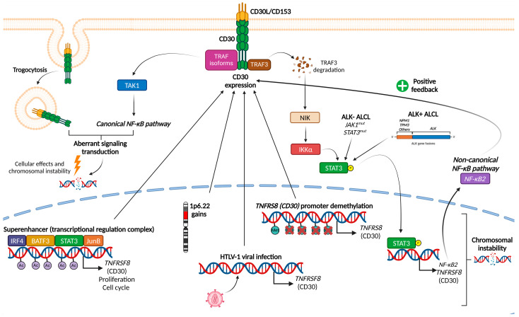
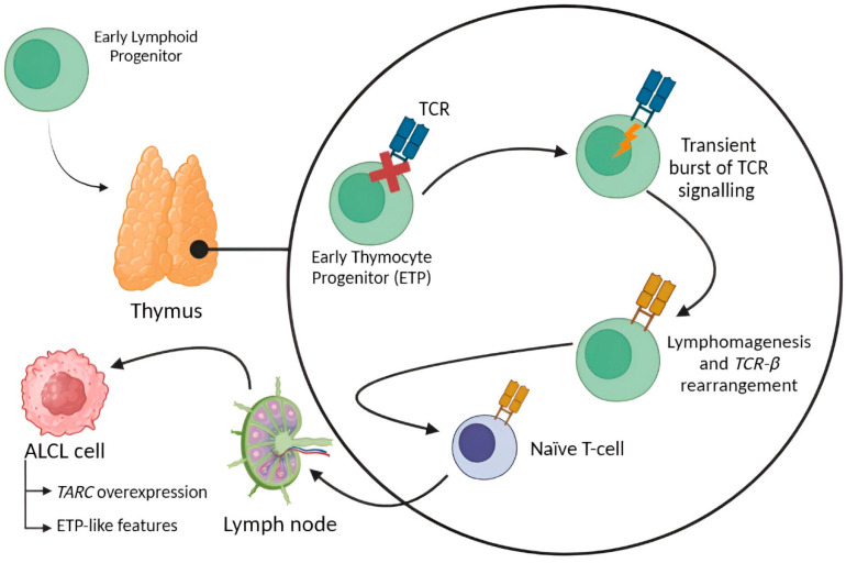
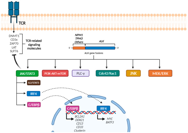
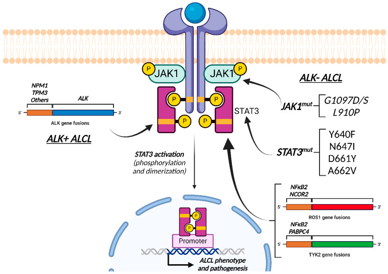
Anaplastic Large Cell Lymphoma (ALCL) represents a diverse group of mature T-Cell Lymphomas unified by strong CD30 expression but with different molecular and clinical subtypes. This review summarizes recent molecular advances in ALCL, highlighting key discoveries that have refined its classification, diagnosis, and therapeutic strategies. ALCL comprises four major entities: systemic ALK-positive ALCL, systemic ALK-negative ALCL, Breast Implant-Associated ALCL (BIA-ALCL), and primary cutaneous ALCL. Each subtype exhibits unique phenotypes, along with cytogenetic and molecular alterations that affect clinical outcomes. Nevertheless, different oncogenic mechanisms mediate STAT3 activation. In ALK-positive ALCL, ALK fusion proteins drive oncogenesis via constitutive activation of STAT3 and other signaling pathways. ALK-negative ALCL comprises heterogeneous genetic subtypes, in which JAK/STAT3 pathway alterations and novel gene fusions are gaining recognition as potential therapeutic targets. This review emphasizes the need for integrative molecular diagnostics to improve stratification of ALCL subtypes and targeted treatment approaches. Future research should focus on elucidating the biological mechanisms underlying these alterations and on translating molecular insights into clinical practice.

Keywords: ALK; Anaplastic Large Cell Lymphoma; BIA-ALCL; DUSP22; STAT3; TP63; pcALCL.


Conflict of interest statement

The authors declare no conflict of interest.

Figures

Figure 1
Figure 1
Different mechanisms leading to CD30 overexpression. Created with BioRender.com.
Figure 2
Figure 2
ALCL etiopathogenesis most accepted theory is supported by TARC overexpression, as well as the presence of ETP-like characteristics. Created with BioRender.com.
Figure 3
Figure 3
Neoplastic small-to-medium-sized cells with homogeneous appearance are partially disrupting lymph node architecture ((A), HE, 10× and (B), HE, 40×). Neoplastic cells express CD30 ((C), 10× and (D), 20×) and ALK with a peculiar exclusive cytoplasmic pattern ((E), 10× and (F), 40×).
Figure 4
Figure 4
Signaling profile of ALK+ ALCL. Created with BioRender.com.
Figure 5
Figure 5
A triple-negative ALCL case for ALK, P63 and DUSP22 translocation is shown. (A) Effacement of the lymph node by a pseudonodular pattern of large atypical cells is seen (HE, 10×). (D) Large atypical cells with different nuclear morphology and size is shown (HE,40×). Neoplastic cells expressed CD30 ((B), 20×), intense nuclear PSTAT3 ((C), 40×), cytoplasmic CD3 ((E), 20×) and perforin ((F), 20×).
Figure 6
Figure 6
A typical case of DUSP22-rearranged ALCL. Numerous “hallmark cells” with “donut-like” nuclear figures are easily found ((A), HE, 40×). Neoplastic cells were pSTAT3 negative ((B), 40×), LEF1 positive ((C), 40×) and TIA1 negative ((D), 20×).
Figure 7
Figure 7
Example of an ALK- ALCL case with JAK2 translocation is shown. (A) Partial effacement of a lymph node by different-sized nodules separated from each others by collagen bundles and inflammatory elements (HE, 4×). (B) At higher-power view, large atypical cells with Hodgkin’s-like looking features intermingled with leukocytes, eosinophils and small lymphocytes are found (HE, 40×). (C) Neoplastic cells express CD30 (4×). (D) Higher-power view of CD30 expression (40×). (E) No PAX5 is expressed on neoplastic cells (20×). (F) Expression of perforin is seen (10×).
Figure 8
Figure 8
Comparison between the different mechanisms for STAT3 activation between ALK+ and ALK- ALCL. Created with BioRender.com.
Figure 9
Figure 9
Example of an ALK- ALCL with p53 overexpression. (A) Complete effacement of lymph node architecture by a diffuse proliferation of large atypical cells (HE staining, 10×). (B) Higher magnification 20×. (C) CD30 expression (20×). (D) Intense p53 expression (20×).
Figure 10
Figure 10
Example of a BIA-ALCL. (A) A breast protesis capsule with fibrinous material in its lumen is shown (HE, 4×). (B) Higher-power view of the fibronous areas show large atypical cells alone or forming small clusters (HE, 40×). (C) Less magnification (2×) and (D) higher magnification (40×) show CD30 expression on neoplastic cells.
Figure 11
Figure 11
Representation of the two main theories established for the development of BIA-ALCL (hypoxia-related and chronic inflammation due to the presence of a bacterial biofilm along the implant). Created with BioRender.com.
Figure 12
Figure 12
Circoplots showing the diverse landscape of gene rearrangements, chromosomal alterations (gains or losses) and SNV mutations observed in different subsets of ALCL. (A) ALK+ ALCL, (B) ALK- ALCL (including BIA-ALCL) and (C) pcALCL.

Similar articles

References

    1. Stansfeld A.G., Diebold J., Noel H., Kapanci Y., Rilke F., Kelényi G., Sundstrom C., Lennert K., van Unnik J.A., Mioduszewska O. Updated Kiel Classification for Lymphomas. Lancet. 1988;1:292–293. doi: 10.1016/S0140-6736(88)90367-4. - DOI - PubMed
    1. Harris N., Jaffe E., Stein H., Banks P., Chan J., Cleary M., Delsol G., De Wolf- Peeters C., Falini B., Gatter K. A Revised European-American Classification of Lymphoid Neoplasms: A Proposal from the International Lymphoma Study Group [See Comments] Blood. 1994;84:1361–1392. doi: 10.1182/blood.V84.5.1361.1361. - DOI - PubMed
    1. Pittaluga S., Wlodarska I., Pulford K., Campo E., Morris S.W., Van den Berghe H., De Wolf-Peeters C. The Monoclonal Antibody ALK1 Identifies a Distinct Morphological Subtype of Anaplastic Large Cell Lymphoma Associated with 2p23/ALK Rearrangements. Am. J. Pathol. 1997;151:343–351. - PMC - PubMed
    1. Pulford K., Lamant L., Morris S.W., Butler L.H., Wood K.M., Stroud D., Delsol G., Mason D.Y. Detection of Anaplastic Lymphoma Kinase (ALK) and Nucleolar Protein Nucleophosmin (NPM)-ALK Proteins in Normal and Neoplastic Cells with the Monoclonal Antibody ALK1. Blood. 1997;89:1394–1404. doi: 10.1182/blood.V89.4.1394. - DOI - PubMed
    1. Duyster J., Bai R.Y., Morris S.W. Translocations Involving Anaplastic Lymphoma Kinase (ALK) Oncogene. 2001;20:5623–5637. doi: 10.1038/sj.onc.1204594. - DOI - PubMed

MeSH terms

Substances

LinkOut - more resources