. 2025 Jun 19;26(12):5877.
doi: 10.3390/ijms26125877.

Antioxidant and Cytotoxic Evaluation of Aqueous Extracts from Hymenochaetaceae Fungi Associated with Endemic Chilean Sclerophyll Forest Trees

Affiliations

Antioxidant and Cytotoxic Evaluation of Aqueous Extracts from Hymenochaetaceae Fungi Associated with Endemic Chilean Sclerophyll Forest Trees

Suleivys M Nuñez et al. Int J Mol Sci. .

Abstract

In the search for safe and effective natural antioxidants, this study investigates the antioxidant and cytotoxic properties of aqueous extracts obtained from three fungi of the family Hymenochaetaceae: Inonotus sp., Fulvifomes sp., and Phylloporia boldo, all associated with endemic trees of the Chilean sclerophyll forest. Antioxidant capacity was assessed through DPPH, ABTS, and hydroxyl radical scavenging assays. Fulvifomes sp. exhibited the highest antioxidant activity across all methods, which was consistent with its elevated polyphenol content. P. boldo, on the other hand, had the highest protein concentration but comparatively lower antioxidant activity. Cytotoxicity was evaluated using the WST-1 assay in the RTgill-W1 salmonid cell line, revealing that Inonotus sp. displayed the lowest cytotoxicity at both tested concentrations, suggesting it may be suitable for bioactive applications in aquaculture. In contrast, Fulvifomes sp. and P. boldo showed significant cytotoxic effects at higher concentrations. These findings highlight the potential of Inonotus sp. as a natural antioxidant with low cytotoxicity and encourages further exploration of native forest fungi as sources of functional bioactive compounds for food, nutraceutical, or aquaculture applications.

Keywords: Fulvifomes sp.; Hymenochaetaceae fungi; Inonotus sp.; Phylloporia boldo; antioxidant activity; aqueous extracts; cytotoxicity; in vitro assays; polyphenols; sclerophyll forest.


Conflict of interest statement

The authors declare no conflicts of interest. The funders had no role in the design of the study; in the collection, analyses, or interpretation of data; in the writing of the manuscript; or in the decision to publish the results.

Figures
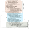
Figure A1
Figure A1
Maximum likelihood tree illustrating a phylogeny of Inonotus, Fulvifomes, and Phylloporia based on ITS sequences. Hymenochaetopsis rigidula was used as outgroup. The maximum likelihood bootstrap values are indicated above the branches (≥70). The sequences generated for the samples Inonotus sp., Fulvifomes sp., and Phylloporia boldo in this research are in bold; the Inonotus clade have pink background, Fulvifomes turquoise background, and Phylloporia grey background.
Figure 1
Figure 1
Concentration of total polyphenols (a) and protein (b) in aqueous extracts of Inonotus sp. (I), Fulvifomes sp. (F), and Phylloporia boldo (Pb). Values are the mean of three replicates ± standard deviation. Different letters indicate significant differences (p ≤ 0.0001) among all groups.
Figure 2
Figure 2
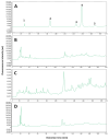
Chromatograms with emission signal obtained at 360 nm of the standard compounds (A) and of the aqueous extracts of P. boldo (B), Fulvifomes sp. (C), and Inonotus sp. (D). The labeled peaks for the chromatogram of the standards are 1. gallic acid, 2. catechin, 3. epicatechin, 4. syringic acid, and 5. p-coumaric acid.
Figure 3
Figure 3
Chromatograms with emission signal obtained at 422 nm of the standard compounds (A) and of the aqueous extracts of P. boldo (B), Fulvifomes sp. (C), and Inonotus sp. (D). The labeled peaks for the chromatogram of the standards are 1. catechin, 2. caffeic acid, 3. epicatechin, 4. syringic acid, and 5. p-coumaric acid.
Figure 4
Figure 4
Fluorescence emission spectrum obtained from the retention time chromatograms of syringic acid. The solid line spectrum is the one extracted from the chromatogram of the syringic acid standard, the one labeled as sample B is the equivalent for the aqueous extract of P. boldo, and sample F is for the extract of Fulvifomes sp.
Figure 5
Figure 5
DPPH scavenging activity of: Inonotus sp. (I), Fulvifomes sp. (F), Phylloporia boldo (Pb), extracts of vitamin C (Vit C) assayed at 3 mg/mL (a,b) Fulvifomes sp. extract, Vit C, Trolox, Gallic acid, black tea with blueberries and maqui (Tea), and the collagen supplement fortified with Vit C and magnesium (Plus) tested at different concentrations. Values are the mean of three replicates ± standard deviation. Different letters indicate significant differences (p ≤ 0.0001) among all groups.
Figure 6
Figure 6
ABTS scavenging activity of: Inonotus sp. (I), Fulvifomes sp. (F), Phylloporia boldo (Pb), extracts of vitamin C (Vit C) assayed at 3 mg/mL (a,b) Fulvifomes sp. extract, Vit C, Trolox, Gallic acid, black tea with blueberries and maqui (Tea), and the collagen supplement fortified with Vit C and magnesium (Plus) tested at different concentrations. Values are the mean of three replicates ± standard deviation. Different letters indicate significant differences (p ≤ 0.0001) among all groups.
Figure 7
Figure 7
Scavenging activity on hydroxyl radicals of: Inonotus sp. (I), Fulvifomes sp. (F), Phylloporia boldo (Pb), extracts of vitamin C (Vit C) assayed at 3 mg/mL (a,b) Fulvifomes sp. extract, Vit C, Trolox, Gallic acid, black tea with blueberries and maqui (Tea) and the collagen supplement fortified with Vit C and magnesium (Plus) tested at different concentrations. Values are the mean of three replicates ± standard deviation. Different letters indicate significant differences (p ≤ 0.0001) among all groups.
Figure 8
Figure 8
Fluorescent microscopy images of fibroblast without extract (control sample) and stained with BCECF-AM (A), propidium iodide (B), Hoechst 33342 (C), or overlap (D) stains. Three separate pictures from the same field were taken for the three markers. Scale bar = 50 μm.
Figure 9
Figure 9
Fluorescent microscopy images of fibroblast treated with aqueous extract (3 mg/mL) of Inonotus sp. and stained with BCECF-AM (A), propidium iodide (B), Hoechst 33342 (C), or overlap (D) stains. Three separate pictures from the same field were taken for the three markers. Scale bar = 50 μm.
Figure 10
Figure 10
Fluorescent microscopy images of fibroblast treated with aqueous extract (3 mg/mL) of Fulviformes spp. and stained with BCECF-AM (A), propidium iodide (B), Hoechst 33342 (C), or overlap (D) stains. Three separate pictures from the same field were taken for the three markers. Scale bar = 50 μm.
Figure 11
Figure 11
Fluorescent microscopy images of fibroblast treated with aqueous extract (3 mg/mL) of P. boldo and stained with BCECF-AM (A), propidium iodide (B), Hoechst 33342 (C), or overlap (D) stains. Three separate pictures from the same field were taken for the three markers. Scale bar = 50 μm.
Figure 12
Figure 12
Viability of RTgill-W1 salmonid cell line evaluated by WST-1 cell proliferation assay after treatments with two different concentrations of Inonotus sp. (I), Fulvifomes sp. (F), or Phylloporia boldo (Pb) aqueous extract. Untreated cells (C(-)) and Triton (T) were performed as negative and positive control, respectively. Values are the mean of three replicates ± standard deviation. Different letters indicate significant differences (p ≤ 0.0001) among all groups.

Similar articles

References

    1. Sies H., Jones D.P. Reactive Oxygen Species (ROS) as Pleiotropic Physiological Signalling Agents. Nat. Rev. Mol. Cell Biol. 2020;21:363–383. doi: 10.1038/s41580-020-0230-3. - DOI - PubMed
    1. Kehm R., Baldensperger T., Raupbach J., Höhn A. Protein Oxidation—Formation Mechanisms, Detection and Relevance as Biomarkers in Human Diseases. Redox Biol. 2021;42:101901. doi: 10.1016/j.redox.2021.101901. - DOI - PMC - PubMed
    1. Berglund H., Jönsson M.T., Penttilä R., Vanha-Majamaa I. The Effects of Burning and Dead-Wood Creation on the Diversity of Pioneer Wood-Inhabiting Fungi in Managed Boreal Spruce Forests. For. Ecol. Manag. 2011;261:1293–1305. doi: 10.1016/j.foreco.2011.01.008. - DOI
    1. Guerra F., Badilla L., Cautín R., Castro M. In Vitro Propagation of Peumus Boldus Mol, a Woody Medicinal Plant Endemic to the Sclerophyllous Forest of Central Chile. Horticulturae. 2023;9:1032. doi: 10.3390/horticulturae9091032. - DOI
    1. Otero C., Klagges C., Morales B., Sotomayor P., Escobar J., Fuentes J.A., Moreno A.A., Llancalahuen F.M., Arratia-Perez R., Gordillo-Fuenzalida F., et al. Anti-Inflammatory Chilean Endemic Plants. Pharmaceutics. 2023;15:897. doi: 10.3390/pharmaceutics15030897. - DOI - PMC - PubMed

LinkOut - more resources