

Retinal Changes in Early-Onset cblC Methylmalonic Acidemia Identified Through Expanded Newborn Screening: Highlights from a Case Study and Literature Review
- PMID: 40565527
- PMCID: PMC12193327
- DOI: 10.3390/genes16060635
Retinal Changes in Early-Onset cblC Methylmalonic Acidemia Identified Through Expanded Newborn Screening: Highlights from a Case Study and Literature Review
Abstract
Background: Methylmalonic acidemia combined with homocystinuria (cblC) can lead to infantile maculopathy. Although significant visual deterioration is commonly reported in early-onset cblC, we found poor awareness regarding formal assessments of ocular complications, especially in newborns, and of how these complications relate to the timing of therapy initiation. In this work, we present our experience and perform a literature review.
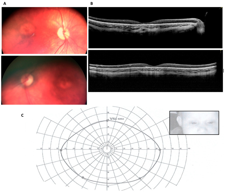
Methods: We performed sequential fundus examinations, optical coherence tomography (OCT) and full-field electroretinography (ERG) under sedation following detection of signs of retinal degeneration. We also assessed visual fields using kinetic attraction perimetry.
Results: We report a newborn who was referred on the eighth day of life, following a diagnosis of cblC through newborn screening (NBS), and who began treatment that same day. Close monitoring of retinal changes through fundus examinations allowed the detection of signs of retinal degeneration at 3 months, which progressed when checked at 5 months. At 7 months, OCT showed retinal thinning with the appearance of bull's eye maculopathy in the corresponding region on fundoscopy; ERG revealed a reduction in the amplitude of both scotopic and photopic components, whereas kinetic attraction perimetry showed no abnormalities. Genetic investigation confirmed the disease, compound heterozygous for a nonsense variant in MMACHC and a splicing one in PRDX1.
Conclusions: In cblC, retinal degeneration occurs in the first months of life despite timely treatment and adequate biochemical control, and it may manifest before any signs of visual deprivation appear. However, there is an early, narrow window during which therapy may slow down retinal degeneration enough to prevent sensory nystagmus. We recommend initiating therapy immediately after biochemical diagnosis, along with close ophthalmological monitoring, before the appearance of any signs.
Keywords: BEM; epi-cblC; infantile maculopathy; motor nystagmus; sensory nystagmus; tandem mass spectrometry biomarkers.
Conflict of interest statement
The authors declare no conflicts of interest.
Figures

Similar articles
-
Variable phenotypes and outcomes associated with the MMACHC c.1A>G variant in Chinese patients with combined methylmalonic acidemia and homocystinuria cblC type.Mol Genet Metab. 2025 Aug;145(4):109182. doi: 10.1016/j.ymgme.2025.109182. Epub 2025 Jun 18. Mol Genet Metab. 2025. PMID: 40544542
-
Optical coherence tomography (OCT) for detection of macular oedema in patients with diabetic retinopathy.Cochrane Database Syst Rev. 2011 Jul 6;(7):CD008081. doi: 10.1002/14651858.CD008081.pub2. Cochrane Database Syst Rev. 2011. Update in: Cochrane Database Syst Rev. 2015 Jan 07;1:CD008081. doi: 10.1002/14651858.CD008081.pub3. PMID: 21735421 Updated.
-
Spectrum of ocular manifestations in cobalamin C and cobalamin A types of methylmalonic acidemia.Ophthalmic Genet. 2016 Dec;37(4):404-414. doi: 10.3109/13816810.2015.1121500. Epub 2016 Mar 15. Ophthalmic Genet. 2016. PMID: 26979128
-
Signs and symptoms to determine if a patient presenting in primary care or hospital outpatient settings has COVID-19.Cochrane Database Syst Rev. 2022 May 20;5(5):CD013665. doi: 10.1002/14651858.CD013665.pub3. Cochrane Database Syst Rev. 2022. PMID: 35593186 Free PMC article.
-
Ocular manifestations in patients with inborn errors of intracellular cobalamin metabolism: a systematic review.Hum Genet. 2022 Jul;141(7):1239-1251. doi: 10.1007/s00439-021-02350-8. Epub 2021 Oct 15. Hum Genet. 2022. PMID: 34652574
References
-
- Ruoppolo M., Malvagia S., Boenzi S., Carducci C., Dionisi-Vici C., Teofoli F., Burlina A., Angeloni A., Aronica T., Bordugo A., et al. Expanded Newborn Screening in Italy Using Tandem Mass Spectrometry: Two Years of National Experience. Int. J. Neonatal Screen. 2022;8:47. doi: 10.3390/ijns8030047. - DOI - PMC - PubMed
-
- Huemer M., Diodato D., Schwahn B., Schiff M., Bandeira A., Benoist J.F., Burlina A., Cerone R., Couce M.L., Garcia-Cazorla A., et al. Guidelines for diagnosis and management of the cobalamin-related remethylation disorders cblC, cblD, cblE, cblF, cblG, cblJ and MTHFR deficiency. J. Inherit. Metab. Dis. 2017;40:21–48. doi: 10.1007/s10545-016-9991-4. - DOI - PMC - PubMed
-
- Guéant J.L., Chéry C., Oussalah A., Nadaf J., Coelho D., Josse T., Flayac J., Robert A., Koscinski I., Gastin I., et al. APRDX1 mutant allele causes a MMACHC secondary epimutation in cblC patients. Nat. Commun. 2018;9:67. doi: 10.1038/s41467-017-02306-5. Erratum in Nat. Commun. 2018, 9, 554. - DOI - PMC - PubMed
-
- Feresin A., Spedicati B., Zampieri S., Morgan A., Magnolato A., Tesser A., Tommasini A., Bonati M.T., Girotto G., Faletra F. Does It Run in Your Family? Inherited Truncating PSMD12 Variants Broaden the Phenotypic Spectrum of Stankiewicz-Isidor Syndrome. Am. J. Med. Genet. Part A. 2024;197:e63953. doi: 10.1002/ajmg.a.63953. - DOI - PubMed
Publication types
MeSH terms
Substances
Supplementary concepts
Grants and funding
LinkOut - more resources
Full Text Sources
Medical
Research Materials
Miscellaneous
