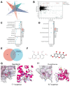
Swertianin Suppresses M1 Macrophage Polarization and Inflammation in Metabolic Dysfunction-Associated Fatty Liver Disease via PPARG Activation
- PMID: 40565585
- PMCID: PMC12193489
- DOI: 10.3390/genes16060693
Swertianin Suppresses M1 Macrophage Polarization and Inflammation in Metabolic Dysfunction-Associated Fatty Liver Disease via PPARG Activation
Abstract
Background: Metabolic dysfunction-associated fatty liver disease (MASLD) is closely associated with immune dysregulation and macrophage-driven inflammation. The activation of PPARG plays a critical role in modulating macrophage polarization and lipid metabolism, suggesting its potential as a therapeutic target for MASLD. Methods: We used UPLC-Q/TOF-MS and network pharmacology to investigate the key components and targets of Swertia davidi Franch, focusing on Swertianin. In vitro experiments on macrophages were conducted to assess the modulation of M1 polarization, and a mouse model of MASLD was utilized to explore the therapeutic effects of Swertianin. Results: Swertianin activated PPARG, leading to significant inhibition of M1 macrophage polarization, a reduction in lipid accumulation, and decreased inflammatory marker levels both in vitro and in vivo. The treatment significantly improved liver pathology in mice, indicating its therapeutic potential for MASLD. Conclusions: Swertianin's activation of PPARG provides a novel mechanism for treating MASLD, targeting both macrophage polarization and inflammation.
Keywords: Swertianin; inflammation; macrophage polarization; metabolic dysfunction-associated fatty liver disease; peroxisome proliferator-activated receptor-gamma.
Conflict of interest statement
The author declares no conflicts of interest.
Figures


Similar articles
-
Hepatoprotective activity of mulberry extract in NAFLD mice for regulating lipid metabolism and inflammation identified via AMPK/PPAR-γ/NF-κB axis.J Ethnopharmacol. 2025 Jul 24;351:120108. doi: 10.1016/j.jep.2025.120108. Epub 2025 Jun 7. J Ethnopharmacol. 2025. PMID: 40490232
-
Phocaeicola dorei ameliorates progression of steatotic liver disease by regulating bile acid, lipid, inflammation and proliferation.Gut Microbes. 2025 Dec;17(1):2539448. doi: 10.1080/19490976.2025.2539448. Epub 2025 Aug 3. Gut Microbes. 2025. PMID: 40754863 Free PMC article.
-
Okra polysaccharide activates PPAR-γ signaling to attenuate metabolic-associated fatty liver disease in mice.Int J Biol Macromol. 2025 Sep;321(Pt 3):146449. doi: 10.1016/j.ijbiomac.2025.146449. Epub 2025 Jul 30. Int J Biol Macromol. 2025. PMID: 40749929
-
Metabolic-dysfunction associated steatotic liver disease and atrial fibrillation: A review of pathogenesis.World J Cardiol. 2025 Jun 26;17(6):106147. doi: 10.4330/wjc.v17.i6.106147. World J Cardiol. 2025. PMID: 40575425 Free PMC article. Review.
-
Comparative efficacy of THR-β agonists, FGF-21 analogues, GLP-1R agonists, GLP-1-based polyagonists, and Pan-PPAR agonists for MASLD: A systematic review and network meta-analysis.Metabolism. 2024 Dec;161:156043. doi: 10.1016/j.metabol.2024.156043. Epub 2024 Sep 30. Metabolism. 2024. PMID: 39357599
References
-
- Pouwels S., Sakran N., Graham Y., Leal A., Pintar T., Yang W., Kassir R., Singhal R., Mahawar K., Ramnarain D. Non-Alcoholic Fatty Liver Disease (NAFLD): A Review of Pathophysiology, Clinical Management and Effects of Weight Loss. BMC Endocr. Disord. 2022;22:63. doi: 10.1186/s12902-022-00980-1. - DOI - PMC - PubMed
MeSH terms
Substances
Grants and funding
- KJZD-K202302701, KJZD-K202202701, KJQN202302725/The Science and Technology Research Program of Chongqing Municipal Education Commission
- sys20210005/The Key laboratory project of Chongqing Three Gorges Medical College
- 2019XZZ002, XJ2021000301, 2023gccrc07/The Natural Science Project of Chongqing Three Gorges Medical College
- CSTB2024NSCQ-MSX0319/Natural Science Foundation of Chongqing
LinkOut - more resources
Full Text Sources
Medical

