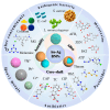
Au-Ag Bimetallic Nanoparticles for Surface-Enhanced Raman Scattering (SERS) Detection of Food Contaminants: A Review
- PMID: 40565718
- PMCID: PMC12192556
- DOI: 10.3390/foods14122109
Au-Ag Bimetallic Nanoparticles for Surface-Enhanced Raman Scattering (SERS) Detection of Food Contaminants: A Review
Abstract
Food contaminants, including harmful microbes, pesticide residues, heavy metals and illegal additives, pose significant public health risks. While traditional detection methods are effective, they are often slow and require complex equipment, which limits their application in real-time monitoring and rapid response. Surface-enhanced Raman scattering (SERS) technology has gained widespread use in related research due to its hypersensitivity, non-destructibility and molecular fingerprinting capabilities. In recent years, Au-Ag bimetallic nanoparticles (Au-Ag BNPs) have emerged as novel SERS substrates, accelerating advancements in SERS detection technology. Au-Ag BNPs can be classified into Au-Ag alloys, Au-Ag core-shells and Au-Ag aggregates, among which the Au-Ag core-shell structure is more widely applied. This review discusses the types, synthesis methods and practical applications of Au-Ag BNPs in food contaminants. The study aims to provide valuable insights into the development of new Au-Ag BNPs and their effective use in detecting common food contaminants. Additionally, this paper explores the challenges and future prospects of SERS technology based on Au-Ag BNPs for pollutant detection, including the development of functional integrated substrates, advancements in intelligent algorithms and the creation of portable on-site detection platforms. These innovations are designed to streamline the detection process and offer guidance in selecting optimal sensing methods for the on-site detection of specific pollutants.
Keywords: Au-Ag bimetallic nanoparticles; Surface-enhanced Raman scattering; food contaminants; sensor.
Conflict of interest statement
The authors declare no conflicts of interest.
Figures


Similar articles
-
Recent Developments and Applications of Surface-Enhanced Raman Scattering Spectroscopy in Pesticides Detection: From Single Pesticides to Mixed Pesticides.ACS Omega. 2025 Jun 12;10(24):25158-25175. doi: 10.1021/acsomega.5c02940. eCollection 2025 Jun 24. ACS Omega. 2025. PMID: 40584312 Free PMC article. Review.
-
A SERS aptasensor based on Au@Ag bimetallic nanostars-magnetic covalent organic composites for rHuEPO-α detection.Anal Chim Acta. 2025 Sep 15;1367:344307. doi: 10.1016/j.aca.2025.344307. Epub 2025 Jun 6. Anal Chim Acta. 2025. PMID: 40610147
-
Multifunctional bionanocomposites for trace detection of water contaminants.J Colloid Interface Sci. 2025 Dec 15;700(Pt 3):138587. doi: 10.1016/j.jcis.2025.138587. Epub 2025 Jul 29. J Colloid Interface Sci. 2025. PMID: 40763505
-
Advances in Hydrogel-Integrated SERS Platforms: Innovations, Applications, Challenges, and Future Prospects in Food Safety Detection.Biosensors (Basel). 2025 Jun 5;15(6):363. doi: 10.3390/bios15060363. Biosensors (Basel). 2025. PMID: 40558445 Free PMC article. Review.
-
Innovative applications of quantum dots-based surface-enhanced Raman spectroscopy for food safety detection.Crit Rev Food Sci Nutr. 2025 Jul 17:1-20. doi: 10.1080/10408398.2025.2531224. Online ahead of print. Crit Rev Food Sci Nutr. 2025. PMID: 40671534 Review.
Cited by
-
Analysis and Optimization of Rotationally Symmetric Au-Ag Alloy Nanoparticles for Refractive Index Sensing Properties Using T-Matrix Method.Nanomaterials (Basel). 2025 Jul 6;15(13):1052. doi: 10.3390/nano15131052. Nanomaterials (Basel). 2025. PMID: 40648759 Free PMC article.
References
-
- Song S.-H., Gao Z.-F., Guo X., Chen G.-H. Aptamer-Based Detection Methodology Studies in Food Safety. Food Anal. Methods. 2019;12:966–990. doi: 10.1007/s12161-019-01437-3. - DOI
-
- Xia X., Zhang B., Wang J., Li B., He K., Zhang X. Rapid Detection of Escherichia coli O157:H7 by Loop-Mediated Isothermal Amplification Coupled with a Lateral Flow Assay Targeting the z3276 Genetic Marker. Food Anal. Methods. 2022;15:908–916. doi: 10.1007/s12161-021-02172-4. - DOI
-
- Zhang Y., Li M., Cui Y., Hong X., Du D. Using of Tyramine Signal Amplification to Improve the Sensitivity of ELISA for Aflatoxin B1 in Edible Oil Samples. Food Anal. Methods. 2018;11:2553–2560. doi: 10.1007/s12161-018-1235-9. - DOI
Publication types
Grants and funding
LinkOut - more resources
Full Text Sources
Miscellaneous

