

Thoracic Ultrasound for Acute Dyspnea in Interstitial Lung Disease
- PMID: 40565904
- PMCID: PMC12194010
- DOI: 10.3390/jcm14124159
Thoracic Ultrasound for Acute Dyspnea in Interstitial Lung Disease
Abstract
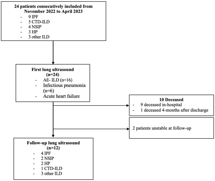
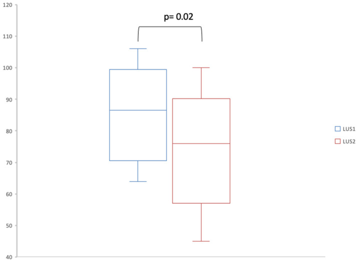
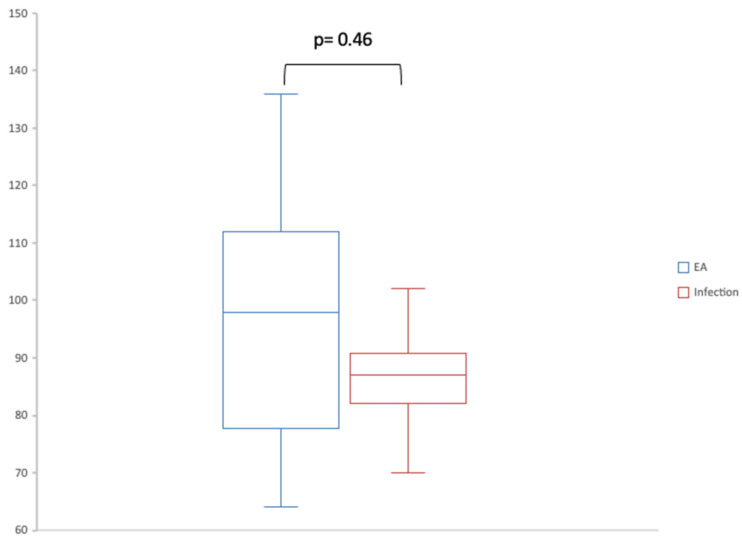
Background: Lung ultrasound (LUS) can be used at follow-up for patients with stable interstitial lung disease (ILD). LUS could also help guide the diagnosis of etiology for acute respiratory episodes. Methods: We conducted a prospective, one-center, observational study including patients with ILD hospitalized in the pulmonology unit or in the intensive care unit of the Tours University Hospital for acute dyspnea. LUS was performed at admission and then at a follow-up visit in the six months following discharge. We compared the number of B-lines between the two LUSs. We also compared the features of the first LUS between the different etiologies responsible for increased dyspnea. Results: Of 24 patients, 16 had acute ILD exacerbation (67%), 6 had pulmonary infections (25%) and 2 had acute heart failure (8%). LUS was feasible in all patients and always showed lung sliding, pleural irregularities and B-lines. There were pleural effusions in four cases (17%) and pulmonary consolidations in two cases (8%). Seven patients had A-lines in at least one thoracic space on the initial LUS. We found a significant decrease in the number of B-lines at follow-up (76; IQR, [59-86.75]) compared to admission (86.5; IQR, [71.5-94.5]) (p-value = 0.02). There was a trend of more A-lines in patients with infection (1 [0.25-1.75]) compared to AE-ILD (0 [0-0]). Conclusions: Following an episode of acute dyspnea in patients with ILD, LUS shows a decrease in the number of B-lines. Patients with ILD and concurrent pulmonary infection may have more A-lines than patients with AE-ILD.
Keywords: dyspnea; exacerbation; infection; interstitial lung disease; lung ultrasound.
Conflict of interest statement
The authors declare no conflicts of interest.
Figures
Similar articles
-
Cyclophosphamide for connective tissue disease-associated interstitial lung disease.Cochrane Database Syst Rev. 2018 Jan 3;1(1):CD010908. doi: 10.1002/14651858.CD010908.pub2. Cochrane Database Syst Rev. 2018. PMID: 29297205 Free PMC article.
-
Pulmonary rehabilitation for interstitial lung disease.Cochrane Database Syst Rev. 2021 Feb 1;2(2):CD006322. doi: 10.1002/14651858.CD006322.pub4. Cochrane Database Syst Rev. 2021. PMID: 34559419 Free PMC article.
-
The diagnostic utility of lung ultrasound in the assessment of interstitial lung disease associated with rheumatoid arthritis.Arthritis Res Ther. 2025 Jul 30;27(1):159. doi: 10.1186/s13075-025-03626-4. Arthritis Res Ther. 2025. PMID: 40739275 Free PMC article.
-
Sertindole for schizophrenia.Cochrane Database Syst Rev. 2005 Jul 20;2005(3):CD001715. doi: 10.1002/14651858.CD001715.pub2. Cochrane Database Syst Rev. 2005. PMID: 16034864 Free PMC article.
-
Signs and symptoms to determine if a patient presenting in primary care or hospital outpatient settings has COVID-19.Cochrane Database Syst Rev. 2022 May 20;5(5):CD013665. doi: 10.1002/14651858.CD013665.pub3. Cochrane Database Syst Rev. 2022. PMID: 35593186 Free PMC article.
References
-
- Cottin V., Crestani B., Cadranel J., Cordier J.F., Marchand-Adam S., Prévot G., Marchand-Adam S., Nunes H., Wémeau-Stervinou L., Bergot E., et al. French practical guidelines for the diagnosis and management of idiopathic pulmonary fibrosis—2017 update. Full-length version. Rev. Mal. Respir. 2017;34:900–968. doi: 10.1016/j.rmr.2017.07.017. - DOI - PubMed
LinkOut - more resources
Full Text Sources
