Review
. 2025 Jun 12;14(12):4166.
doi: 10.3390/jcm14124166.

Fibromyalgia in the Era of Brain PET/CT Imaging

Affiliations
Review

Fibromyalgia in the Era of Brain PET/CT Imaging

Elisabetta Abenavoli et al. J Clin Med. .

Abstract

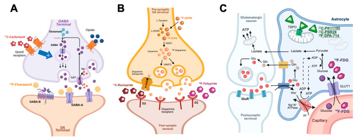
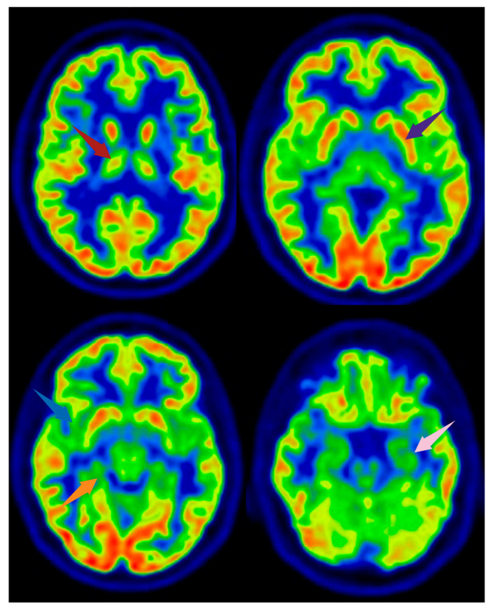
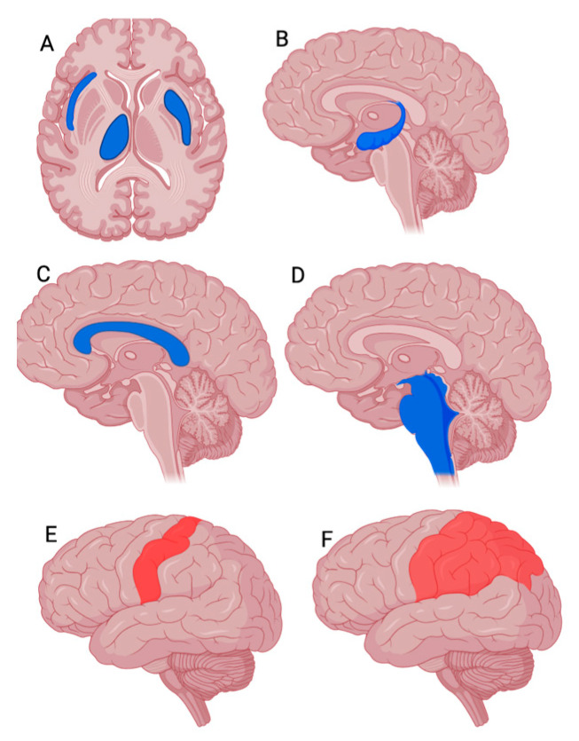
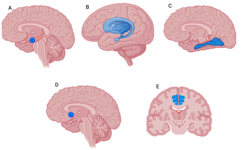
Fibromyalgia syndrome (FMS) is a complex, heterogeneous disorder characterized by chronic widespread pain, fatigue, and cognitive disturbances. The multifactorial nature of FMS, with the involvement of central and peripheral mechanisms, hampers diagnosis and effective treatment. In recent years, positron emission tomography (PET) imaging has emerged as a valuable tool for exploring the neurobiological underpinnings of FMS. Several studies have investigated alterations in glucose metabolism, neurotransmitter systems (including opioid, dopamine, and GABAergic pathways), and neuroinflammation using various PET tracers. These findings have revealed distinct brain metabolic and molecular patterns in FMS patients compared to healthy controls, particularly in pain-related regions such as the thalamus, insula, and anterior cingulate cortex (ACC). Moreover, preliminary data suggest that PET imaging may help identify FMS subgroups with different pathophysiological profiles, potentially allowing for tailored therapeutic approaches. This review summarizes the current evidence on PET applications in FMS and discusses the potential role of molecular imaging in improving patient stratification and predicting treatment response.

Keywords: PET/CT; brain; central nervous system; fibromyalgia syndrome.


Conflict of interest statement

The authors declare no conflicts of interest.

Figures

Figure 1
Figure 1
Principal PET radiopharmaceuticals and their targets investigated in FMS. (A) GABAergic synapses: [11C]Carfentanil targeting pre-synaptic opioid receptors, and [18F]Flumazenil targeting post-synaptic GABAA receptors; (B) Dopaminergic synapses: [18F]DOPA targeting DA synthesis, and [11C]Raclopride or [18F]Fallypride targeting post-synaptic D2 receptors; (C) Glutamatergic synapses: [18F]FDG targeting glucose metabolism and synaptic function, and [11C]PK11195 targeting TSPO receptors. GABA: gamma-amino-butyric acid; GAT: GABA transporter; DA: dopamine/dopaminergic; D2: Dopamine receptor 2; VMAT: vesicular monoamine transporter; AADC: aromatic L-amino acid decarboxylase; [18F]DOPA: [18F]dihydroxyphenylalanine; [18F]FDG: [18F]fluorodeoxyglucose; GLUT: glucose transporter; Glu: glutamate; Gln: glutamine; ATP: adenosine triphosphate; ADP: adenosine diphosphate; GluR: glutamate receptor. Created using BioRender.com.
Figure 2
Figure 2
[18F]FDG PET images from a healthy subject. Arrows indicate brain regions commonly reported as altered in FMS, shown here for illustrative purposes. The subject depicted did not have FMS. Axial slices are shown. Red arrow: right thalamus; purple arrow: left lentiform nucleus; blue arrow: right insula; orange arrow: parahippocampal gyrus; pink arrow: medial temporal cortex. Image created using BioRender.com.
Figure 3
Figure 3
Representative PET findings in FMS patients compared to healthy controls. Created using BioRender.com. (A,B) [18F]FDG PET images (axial and sagittal slices): regions in blue indicate reduced glucose metabolism in the right thalamus, right insula, and left striatum (axial), and hippocampus (sagittal) in FMS patients. (C,D) [18F]DOPAPET images (sagittal slices): blue regions represent decreased presynaptic dopaminergic activity in the limbic system and brainstem. (E,F) TSPO PET images (e.g., [11C]PBR28 or [18F]DPA-714; sagittal slices): red regions indicate increased tracer uptake reflecting neuroinflammation in the primary somatosensory cortex and right parietal gray matter. Color scale interpretation: blue areas indicate reduced tracer uptake in FMS patients compared to healthy controls; red areas indicate increased uptake.
Figure 4
Figure 4
Representative PET findings in FMS patients compared to healthy controls. Created using BioRender.com. (A) [18F]FDG PET sagittal slice. Blue regions indicate reduced glucose metabolism in the amygdala of FMS patients compared to healthy controls. (B) [11C]Raclopride PET sagittal slice. Blue regions reflect decreased dopaminergic receptor availability in the basal ganglia. (C) [18F]Fallypride PET sagittal slice. Blue regions show reduced D2/D3 receptor binding in the fusiform gyrus. (D) [11C]Carfentanil PET sagittal slice. Blue regions represent lower MOR availability in the nucleus accumbens. (E) [11C]PK11195 PET coronal slice. Blue regions demonstrate reduced TSPO binding in the ACC of FMS patients. Color coding: Blue = reduced radiotracer uptake or receptor availability in FMS patients compared to healthy control.

Similar articles

References

    1. Al Sharie S., Varga S.J., Al-Husinat L., Sarzi-Puttini P., Araydah M., Bal’awi B.R., Varrassi G. Unraveling the Complex Web of Fibromyalgia: A Narrative Review. Medicina. 2024;60:272. doi: 10.3390/medicina60020272. - DOI - PMC - PubMed
    1. Sarzi-Puttini P., Giorgi V., Marotto D., Atzeni F. Fibromyalgia: An update on clinical characteristics, aetiopathogenesis and treatment. Nat. Rev. Rheumatol. 2020;16:645–660. doi: 10.1038/s41584-020-00506-w. - DOI - PubMed
    1. Varrassi G., Rekatsina M., Perrot S., Bouajina E., Paladini A., Coaccioli S., Narvaez Tamayo M.A., Sarzi Puttini P. Is Fibromyalgia a Fashionable Diagnosis or a Medical Mystery? Cureus. 2023;15:e44852. doi: 10.7759/cureus.44852. - DOI - PMC - PubMed
    1. Fitzcharles M.A., Yunus M.B. The clinical concept of fibromyalgia as a changing paradigm in the past 20 years. Pain Res. Treat. 2012;2012:184835. doi: 10.1155/2012/184835. - DOI - PMC - PubMed
    1. Arout C.A., Sofuoglu M., Bastian L.A., Rosenheck R.A. Gender Differences in the Prevalence of Fibromyalgia and in Concomitant Medical and Psychiatric Disorders: A National Veterans Health Administration Study. J. Womens Health. 2018;27:1035–1044. doi: 10.1089/jwh.2017.6622. - DOI - PMC - PubMed

LinkOut - more resources