. 2025 Jun 12;14(12):4198.
doi: 10.3390/jcm14124198.

Pyodermatitis-Pyostomatitis Vegetans: The Role of Langerin Deficiency in Disease Pathogenesis

Affiliations

Pyodermatitis-Pyostomatitis Vegetans: The Role of Langerin Deficiency in Disease Pathogenesis

Dan Pan et al. J Clin Med. .

Abstract

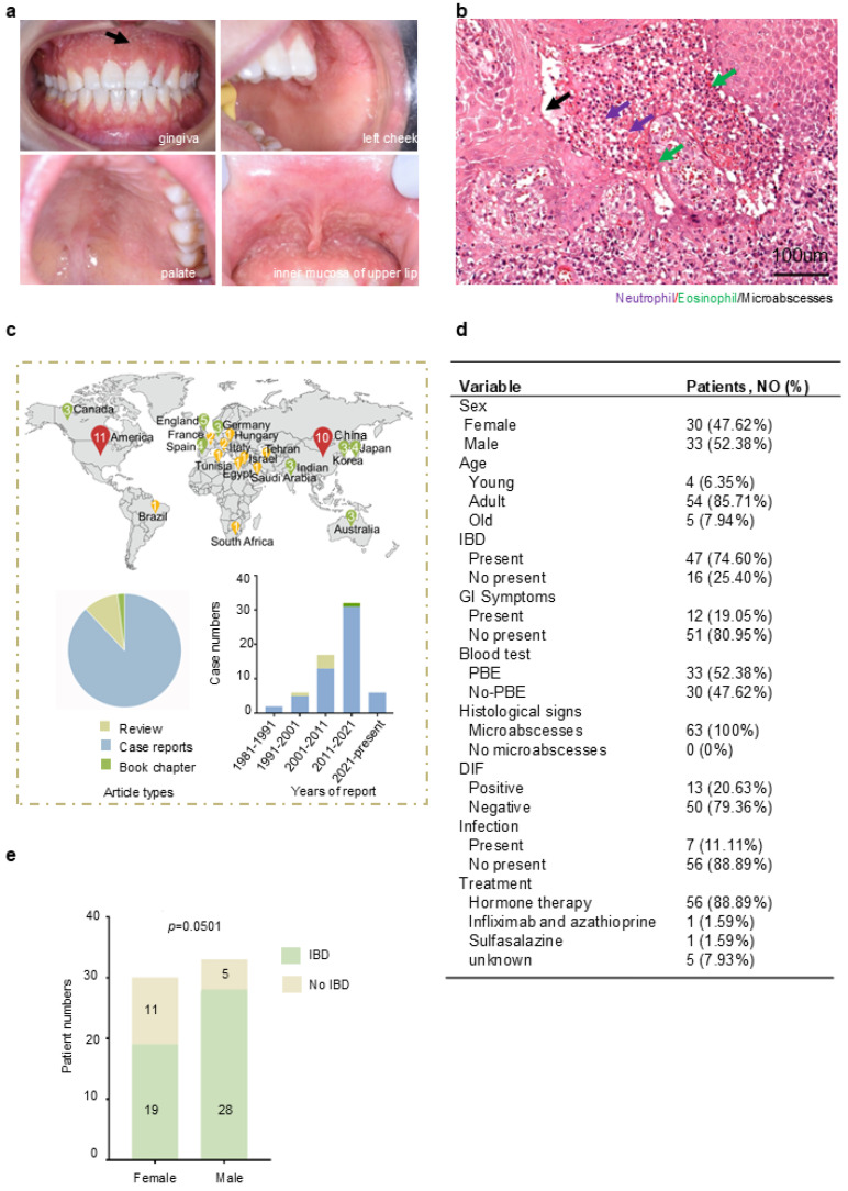
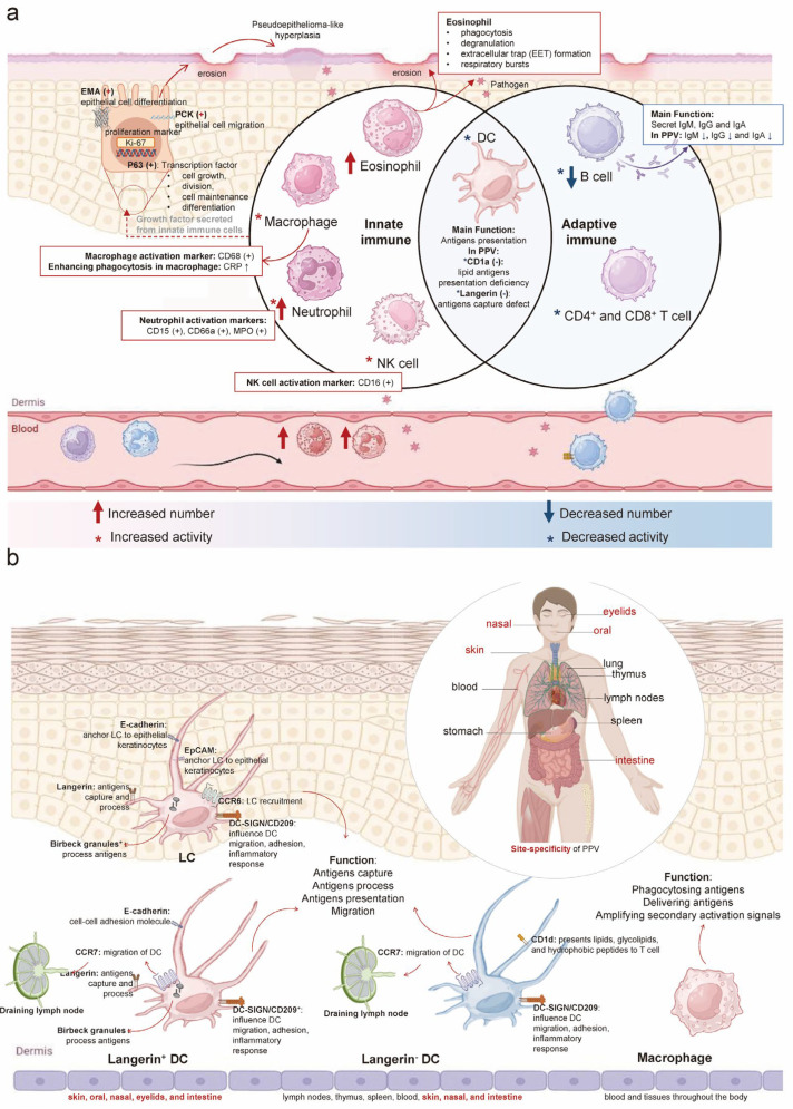
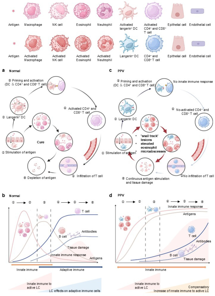
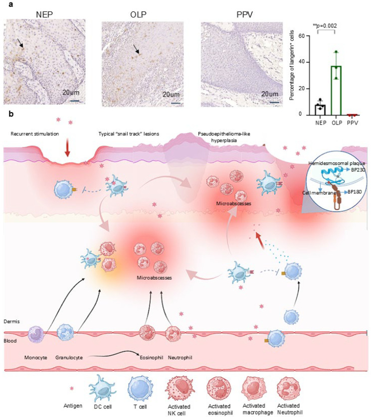
Background/Objectives: Pyodermatitis-pyostomatitis vegetans (PPV) is a rare, chronic inflammatory mucocutaneous disorder. However, the etiology of PPV remains controversial. Methods: A review of online PPV case studies from PubMed, Wanfang database, Web of Science, and books has been performed. Comparative analysis of langerin expression has been conducted to verify the hypothesis summarized from the literature review by Immunohistochemistry (IHC). Results: A total of 63 patients were analyzed across 5 reviews, 44 case reports, and 1 book chapter. Our findings revealed distinct immunological alterations in PPV patients. Innate immunity was upregulated, marked by increased neutrophil and eosinophil counts and enhanced macrophage activity. Adaptive immunity was suppressed, with reduced dendritic cell (DC) numbers and activity and diminished adaptive immune responses. We hypothesize that langerin was a critical factor, contributing to adaptive immune suppression and a compensatory innate immune hyperactivation. Conclusions: We propose the hypothesis that langerin expression on Langerhans cells (LCs) plays a pivotal role in PPV pathogenesis by shifting the immune balance toward innate hyperactivation at the expense of adaptive immunity.

Keywords: Pyodermatitis–pyostomatitis vegetans; adaptive immune; dendritic cells; innate immune; langerin.


Conflict of interest statement

The authors declare no conflicts of interest.

Figures

Figure 1
Figure 1
Reveal typical clinical and histological features of PPV: (a): clinical manifestation, extensive yellowish-white erosions, formation of pseudoepitheliomatous hyperplasia, and “snail track” lesions (black arrow) on the gingiva, left cheek, palate, and inner mucosa of upper lip. (b): Histological manifestation (H&E stain, original magnification ×200 μm); Infiltration of neutrophil (purple arrows) and eosinophil (green arrows) formed microabscesses (black arrows). One patient with PSV in the West China School of Stomatology, Sichuan University. (c): The distribution of PPV in different countries was shown in map, and the number inside the icon indicates the number of cases reported in that country. Article types of PPV cases and years of case reports were summarized. (d): Summary of 63 cases’ clinic features in PPV. (e): Analysis of sex difference and concomitant with IBD in PPV (p = 0.0501). Additional data are in (Supplementary Figure S1). Data are presented as mean ± S.E.M. Group differences were analyzed with the chi-square test.
Figure 2
Figure 2
Langerin deficiency results in enhancement of innate immunity and weakening of adaptive immunity in PPV: (a): In PPV, innate immune cells such as eosinophils, macrophages, neutrophils, NK cells, and DC show increased numbers and/or activity, except for DC. Adaptive immune cells, including DC, B cells, and T cells, exhibit decreased numbers and activity. CD68, a marker of macrophages, is positive along with increased CRP, enhancing macrophage phagocytosis and indicating increased macrophage activity. CD15, CD66a, and MPO are positive as neutrophil markers, reflecting neutrophil activation. CD16 positivity indicates NK cell activation. CD1a and langerin are negative, both considered DC markers. A reduction in IgA, IgM, and IgG antibodies produced by B cells in serum suggests decreased B cell activity and numbers in PPV. Positive PCK (regulating cell migration), EMA (regulating epithelial cell differentiation), P63 (regulating cell proliferation), and Ki-67 (cell proliferation marker) are observed in response to recurrent erosion and epithelial hyperplasia lesions of PPV on the skin and mucosa. (b): Four types of APCs are found in humans: LC, langerin+ DC, langerin DC, and macrophage. LC and langerin+ DCs are distributed in the skin, oral cavity, nasal passages, eyelids, and intestines. Langerin DCs are found in lymph nodes, thymus, spleen, blood, skin, nasal passages, and intestines. Macrophages are distributed throughout the blood and tissues of the body. LC, langerin+ DC, and langerin DC share functions like antigen capture, processing, presentation, and migration. Besides phagocytosing particulate antigens, macrophages enhance the immune response by delivering antigens and amplifying secondary activation signals. The expression of langerin, CD1a, Birbeck granules, E-Cadherin, Epithelial-cell adhesion molecule (EpCAM), CC-chemokine receptor 6 (CCR6), CC-chemokine receptor 7 (CCR7), CD1d, and DC-SIGN shown in four types of APCs. (CRP: C-reactive protein; MPO: Myeloperoxidase; PCK: Pan cytokeratin; EMA: Epithelial membrane antigens; APCs: Antigen-presenting cells).
Figure 3
Figure 3
Immune response to antigens in NEP and enhancement of innate immunity in response to langerin deficiency in PPV: (a) Under normal circumstances, innate immune cells, including macrophages, NK cells, eosinophils, and neutrophils, serve as the first line of defense against pathogen invasion, prompting the adaptive immune process. Antigens are internalized into Birbeck granules in DC for processing and presented to T cells. Following this, CD4+ and CD8+ T cell is activated, marking the onset of adaptive immunity and T cell infiltration. The antibodies secreted by B cells, along with the effector and cytotoxic responses released by T cells, eliminate antigens and maintain a normal physiological state. (b): As the adaptive immune response strengthens, the role of the innate immune system gradually diminishes, and pathogens are progressively eliminated. The pathogen load peaks during antigen presentation and gradually decreases until it disappears with the activation of adaptive immunity. In the early stages of the immune response, tissue damage gradually increases, but with the advent of adaptive immunity, tissue damage is repaired. (c): In the case of PPV, due to the lack of langerin, DCs fail to present antigens to CD4+ and CD8+ T cells. This failure interrupts T cell activation and infiltration, leading to decreased antibody secretion by B cells and reduced effector and cytotoxic responses by T cells. (d): Concurrently, the weakened adaptive immunity prompts a compensatory enhancement of the innate immune response, evidenced by increased macrophage, eosinophil, and neutrophil. Due to the weakening of adaptive immunity in PPV, antigens present a stronger response from the innate immune system. The persistent antigens cannot be eliminated due to the dysfunction of the adaptive immune system, leading to accumulated tissue damage.
Figure 4
Figure 4
Identification of langerin negative in PPV interprets the clinicopathological manifestations of PPV: (a): Four NEP, three OLP, and two PPV tissues were used to stain langerin. The langerin expression in PPV was barely compared to NEP, with an 8% decrease. Langerin expression was increased by about 29% in patients with OLP compared with NEP (** p = 0.002, 29.330 ± 1.156). The expression of langerin in PPV was reduced by about 30% compared with that in OLP. Scale bar = 20 μm. Data are presented as mean ± S.E.M.; ns: no statistical significance; ** p ≤ 0.01. Group differences were analyzed with Student’s unpaired tests. (PPV: Pyodermatitis–pyostomatitis vegetans; NEP: Normal epithelium; OLP: Oral lichen planus). (b): Antigens cross epithelial and endothelial cells to stimulate the innate immune system. The lack of langerin impairs adaptive immune function, leading to a compensatory increase in innate immunity. Neutrophil migrates from the bloodstream to lesion sites, where the infiltration of eosinophils and neutrophils forms microabscesses. Recurrent stimulation by inflammatory factors and antigens leads to the formation of damaged epithelial cells, clinically presenting as nodular hyperplasia and pseudoepitheliomatous hyperplasia. Additionally, the recurrent cycle of lesion stimulation and healing, along with the fusion of damaged and adjacent normal epithelial cells, results in the characteristic “snail track” lesions in PPV. Due to epithelial inflammation, damage to the basal layer, BP180 and BP230 antigens in basement membrane area are exposed to dermis, and BP180 and BP230 antibodies are produced as a result to produce.

Similar articles

References

    1. Gheisari M., Zerehpoosh F.B., Zaresharifi S. Pyodermatitis-pyostomatitis vegetans: A case report and review of literature. Dermatol. Online J. 2020;26:5. doi: 10.5070/D3265048779. - DOI - PubMed
    1. Fde A.M., Rosa D.J., Carvalho M.T., Castañon M.C. Pyodermatitis-pyostomatitis vegetans: Case report and review of medical literature. An. Bras. Dermatol. 2011;86:S137–S140. - PubMed
    1. De Giorgi V., Trane L., Silvestri F., Venturi F., Zuccaro B., Scarfì F. Pyodermatitis-Pyostomatitis Vegetans in Active Inflammatory Bowel Disease. Am. J. Gastroenterol. 2021;116:2163. doi: 10.14309/ajg.0000000000001324. - DOI - PubMed
    1. Ficarra G., Baroni G., Massi D. Pyostomatitis vegetans: Cellular immune profile and expression of IL-6, IL-8 and TNF-alpha. Head Neck Pathol. 2010;4:1–9. doi: 10.1007/s12105-009-0149-7. - DOI - PMC - PubMed
    1. Ahn B.K., Kim S.C. Pyodermatitis-pyostomatitis vegetans with circulating autoantibodies to bullous pemphigoid antigen 230. J. Am. Acad. Dermatol. 2004;50:785–788. doi: 10.1016/j.jaad.2003.09.007. - DOI - PubMed

LinkOut - more resources