. 2025 Jun 15;14(12):4250.
doi: 10.3390/jcm14124250.

Volume Change Measurements of the Heart and Lungs After Pectus Excavatum Repair

Affiliations

Volume Change Measurements of the Heart and Lungs After Pectus Excavatum Repair

Gongmin Rim et al. J Clin Med. .

Abstract

Background/Objectives: The primary objective of PE repair is to relieve compression exerted on the cardiac and pulmonary structures and enhance the thoracic cavity volume. However, the number of volumetric studies of the thoracic cavity, including the heart and lung volumes, is scarce. This study seeks to systematically evaluate the volumetric changes in these structures to assess the physiological impact obtained by PE repair. Methods: A retrospective analysis was conducted on 63 patients who underwent PE repair using the XI bar technique from April 2023 to February 2024. Volumetric changes were measured preoperatively and postoperatively using SYNAPSE 3D imaging software (Version 4.6, Fujifilm, Tokyo, Japan). Cardiac and pulmonary volumes were quantified, and CT indexes (Haller index, Depression index) were assessed. Complication rates, reoperation rates, and length of hospital stay were also analyzed. Results: The mean cardiac volume increased significantly from 458.25 mL preoperatively to 499.13 mL postoperatively (p = 0.018), showing an 8.9% increase. Pulmonary volumes, however, showed no statistically significant change, remaining stable at approximately 4371.31 mL preoperatively and 4266.87 mL postoperatively (p = 0.57). Conclusions: Repairing PE markedly enhances cardiac volume, emphasizing its importance in relieving mediastinal compression. Pulmonary volumes remain largely unaffected, suggesting that PE primarily impacts cardiac structures. Our approach to the volumetric measurements provides valuable insights into the physiological outcomes of chest wall remodeling and is considered to be a good modality for future studies to enhance our understanding of the functional benefits of PE repair.

Keywords: Pectus excavatum; XI technique; cardiac volume; volumetric measurement.


Conflict of interest statement

The authors declare no conflicts of interest.

Figures
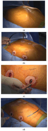
Figure 1
Figure 1
The four steps involved in crane-powered entire chest wall remodeling were as follows: (a) Total elevation of the sternum using a screw crane to exceed the desired chest wall remodeling level. (b) Introduction of multiple pectus bars at the bilateral anterior axillary lines in an XI configuration for comprehensive chest wall remodeling. (c) Stabilization of pectus bars with bilateral bridge plates, linking the bars using nuts and bolts at the subcutaneous plane. (d) Correction of lower costal flares and residual protrusions using the Flare-buster and Magic string technique with heavy strings.
Figure 2
Figure 2
Three-dimensional reconstruction of the patient’s chest CT scan using SYNAPSE 3D: (a) Preoperative images. (b) Postoperative images following XI repair. (c) Three-dimensional reconstructed images of the heart and lungs before the repair. (d) Three-dimensional reconstructed images of the heart and lungs after the repair.
Figure 2
Figure 2
Three-dimensional reconstruction of the patient’s chest CT scan using SYNAPSE 3D: (a) Preoperative images. (b) Postoperative images following XI repair. (c) Three-dimensional reconstructed images of the heart and lungs before the repair. (d) Three-dimensional reconstructed images of the heart and lungs after the repair.
Figure 3
Figure 3
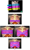
Three zones of chest wall deformities and coverage areas of different pectus bar configurations. (a) Zone 1: deepest point of the sternum; Zone 2: upper chest wall deformity; Zone 3: lateral chest wall and lower costal flares. (b) Coverage area of a single pectus bar, primarily addressing Zone 1. (c) Coverage area of double pectus bars: parallel-shaped bars addressing Zones 1 and 2, and cross-shaped bars covering Zones 1 and 3. (d) Coverage area of the XI pectus bar, comprehensively addressing Zones 1, 2, and 3.

Similar articles

References

    1. Janssen N., Coorens N.A., Franssen A.J., Daemen J.H., Michels I.L., Hulsewé K.W., Vissers Y.L., de Loos E.R. Pectus excavatum and carinatum: A narrative review of epidemiology, etiopathogenesis, clinical features, and classification. J. Thorac. Dis. 2024;16:1687–1701. doi: 10.21037/jtd-23-957. - DOI - PMC - PubMed
    1. Choi J.H., Park I.K., Kim Y.T., Kim W.S., Kang C.H. Classification of Pectus Excavatum According to Objective Parameters From Chest Computed Tomography. Ann. Thorac. Surg. 2016;102:1886–1891. doi: 10.1016/j.athoracsur.2016.05.079. - DOI - PubMed
    1. Park H.J., Lee S.Y., Lee C.S., Youm W., Lee K.R. The Nuss procedure for pectus excavatum: Evolution of techniques and early results on 322 patients. Ann. Thorac. Surg. 2004;77:289–295. doi: 10.1016/S0003-4975(03)01330-4. - DOI - PubMed
    1. Park H.J., Rim G., Yoon S.K. Recent Advancements in Pectus Surgery: Crane Lifting, Multiple-Bar Approach, Bridge Stabilization, and Sandwich Technique. J. Pediatr. Surg. 2024;59:640–647. doi: 10.1016/j.jpedsurg.2023.12.012. - DOI - PubMed
    1. Park H.J., Jeong J.Y., Jo W.M., Shin J.S., Lee I.S., Kim K.T., Choi Y.H. Minimally invasive repair of pectus excavatum: A novel morphology-tailored, patient-specific approach. J. Thorac. Cardiovasc. Surg. 2010;139:379–386. doi: 10.1016/j.jtcvs.2009.09.003. - DOI - PubMed

LinkOut - more resources