

Changes of Airway Space and Flow in Patients Treated with Rapid Palatal Expander (RPE): An Observational Pilot Study with Comparison with Non-Treated Patients
- PMID: 40566102
- PMCID: PMC12194650
- DOI: 10.3390/jcm14124357
Changes of Airway Space and Flow in Patients Treated with Rapid Palatal Expander (RPE): An Observational Pilot Study with Comparison with Non-Treated Patients
Abstract
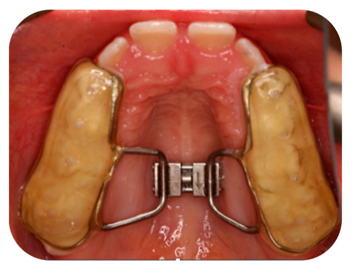
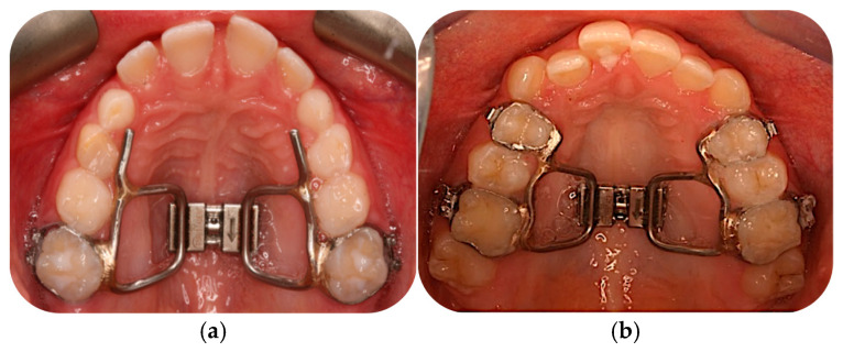
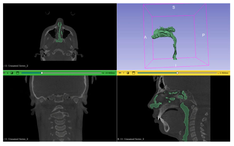
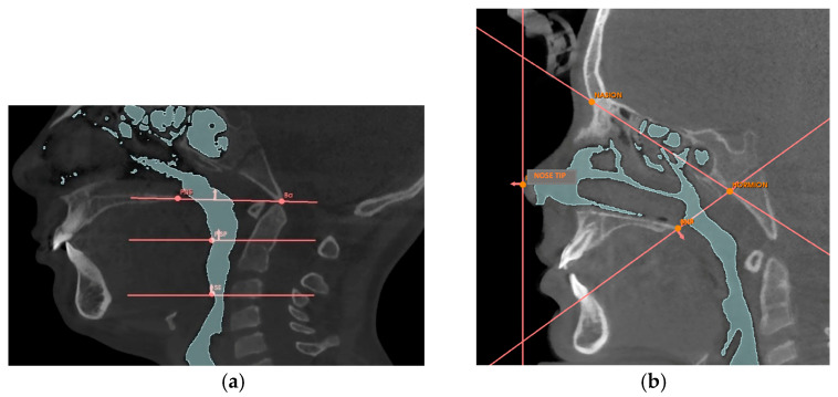
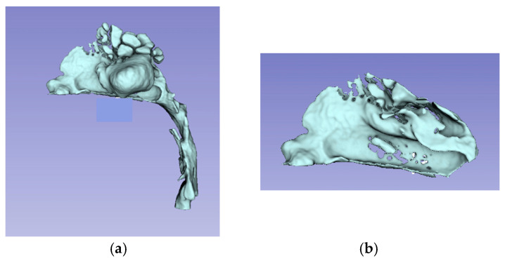
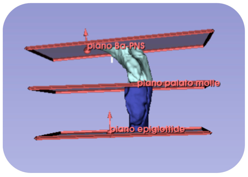
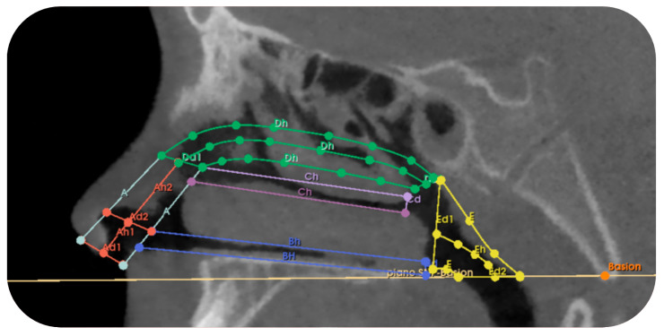
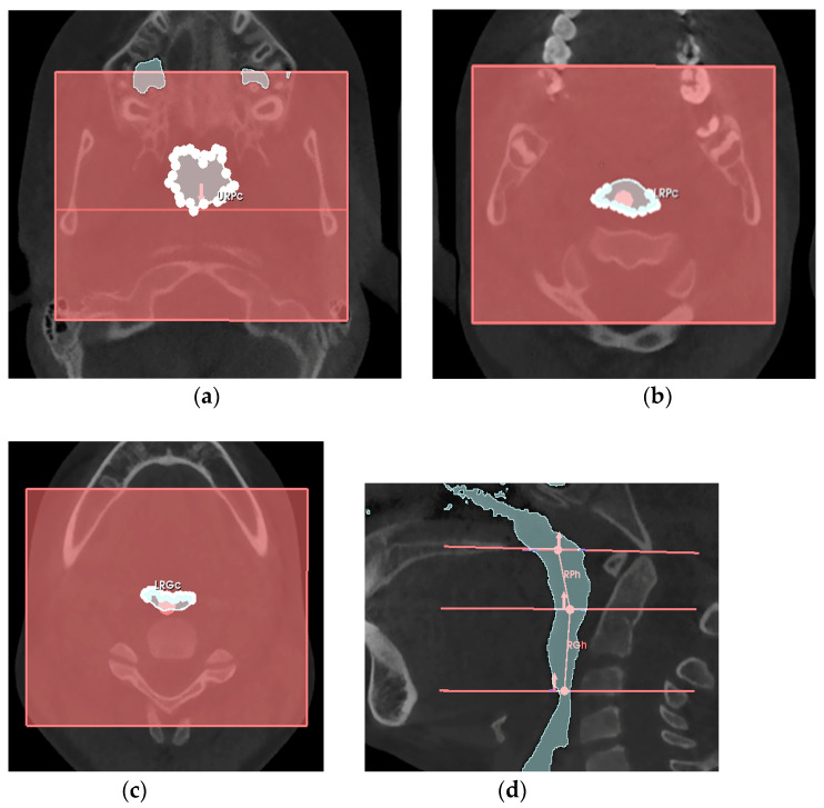
Background/Objectives. With a rapid palatal expander (RPE) is reported to be effective in increasing the volume of nasal cavities, with a restoration of physiological nasal airflow. The purpose of this retrospective clinical study was to evaluate, using Cone Beam Computed Tomography (CBCT), the volumetric changes and airflow velocity changes in the nasal cavities, retro-palatal and retro-glossal airways, resulting from the use of RPE with dental anchorage (group A), also comparing these data with patients non treated with RPE (group B). Methods. Sixteen subjects (aged 9.34 years) with transverse maxillary deficiency and unilateral posterior crossbite were treated with RPE with dental anchorage. Additionally, 8 patients (aged 11.11 years) with juvenile idiopathic arthritis, who did not undergo any orthodontic treatment, were selected as a control group. Expansion was performed until overcorrection was achieved, and the device was left in place for 6 months as fixed retention, followed by another 6 months of night-time removable retention. From the retrospective evaluation, all patients presented two CBCT scans at baseline (T0) and 1-year follow-up (T1). The 3D-Slicer software was used for each CBCT to measure the nasal (VN), retropalatal (VRP), and retroglossal (VRG) volumes, while an iterative Excel spreadsheet allowed for a pilot approximated modeling and calculation of airway flow-related data. Results. Regarding mean age, a statistically significant difference (p = 0.01 *) was found between groups, suggesting that group B is closer to the pubertal growth peak. Analysis between T0 and T1 revealed: (i) a statistically significant increase for volumes VN, VRP and VRG in group A; (ii) a statistically significant increase for VN in group B; (iii) a statistically significant decrease for all variables related to airflow velocity in both groups. Furthermore, comparison between group A and B, regarding variations between T0 and T1, found a statistically significant difference only for VN. Conclusions. Within the limitations of this pilot evaluation, the treatment with RPE revealed promising outcomes for retro-palatal, retro-glossal and nasal volumes, together with clinical changes in airflow velocities.
Keywords: airway flow; airway volume; cone beam computed tomography; rapid palatal maxillary expansion.
Conflict of interest statement
The authors declare no conflicts of interest.
Figures
Similar articles
-
Sertindole for schizophrenia.Cochrane Database Syst Rev. 2005 Jul 20;2005(3):CD001715. doi: 10.1002/14651858.CD001715.pub2. Cochrane Database Syst Rev. 2005. PMID: 16034864 Free PMC article.
-
Orthodontic treatment for posterior crossbites.Cochrane Database Syst Rev. 2001;(1):CD000979. doi: 10.1002/14651858.CD000979. Cochrane Database Syst Rev. 2001. Update in: Cochrane Database Syst Rev. 2014 Aug 08;(8):CD000979. doi: 10.1002/14651858.CD000979.pub2. PMID: 11279699 Updated.
-
Systemic pharmacological treatments for chronic plaque psoriasis: a network meta-analysis.Cochrane Database Syst Rev. 2021 Apr 19;4(4):CD011535. doi: 10.1002/14651858.CD011535.pub4. Cochrane Database Syst Rev. 2021. Update in: Cochrane Database Syst Rev. 2022 May 23;5:CD011535. doi: 10.1002/14651858.CD011535.pub5. PMID: 33871055 Free PMC article. Updated.
-
Systemic pharmacological treatments for chronic plaque psoriasis: a network meta-analysis.Cochrane Database Syst Rev. 2017 Dec 22;12(12):CD011535. doi: 10.1002/14651858.CD011535.pub2. Cochrane Database Syst Rev. 2017. Update in: Cochrane Database Syst Rev. 2020 Jan 9;1:CD011535. doi: 10.1002/14651858.CD011535.pub3. PMID: 29271481 Free PMC article. Updated.
-
Does Augmenting Irradiated Autografts With Free Vascularized Fibula Graft in Patients With Bone Loss From a Malignant Tumor Achieve Union, Function, and Complication Rate Comparably to Patients Without Bone Loss and Augmentation When Reconstructing Intercalary Resections in the Lower Extremity?Clin Orthop Relat Res. 2025 Jun 26. doi: 10.1097/CORR.0000000000003599. Online ahead of print. Clin Orthop Relat Res. 2025. PMID: 40569278
References
-
- Calvo-Henriquez C., Capasso R., Chiesa-Estomba C., Liu S.Y., Martins-Neves S., Castedo E., O’Connor-Reina C., Ruano-Ravina A., Kahn S. The role of pediatric maxillary expansion on nasal breathing. A systematic review and meta-analysis. Int. J. Pediatr. Otorhinolaryngol. 2020;135:110139. doi: 10.1016/j.ijporl.2020.110139. - DOI - PubMed
-
- Petré S., Bondemark L., Björk O., Derfeldt S. A systematic review concerning early orthodontic treatment of unilateral posterior crossbite. Angle Orthod. 2003;73:588–595. - PubMed
LinkOut - more resources
Full Text Sources
