

Histological and Immunohistochemical Insights into Disc Perforation in the Temporomandibular Joint: A Case Report
- PMID: 40566404
- PMCID: PMC12015778
- DOI: 10.3390/jfmk10020107
Histological and Immunohistochemical Insights into Disc Perforation in the Temporomandibular Joint: A Case Report
Abstract
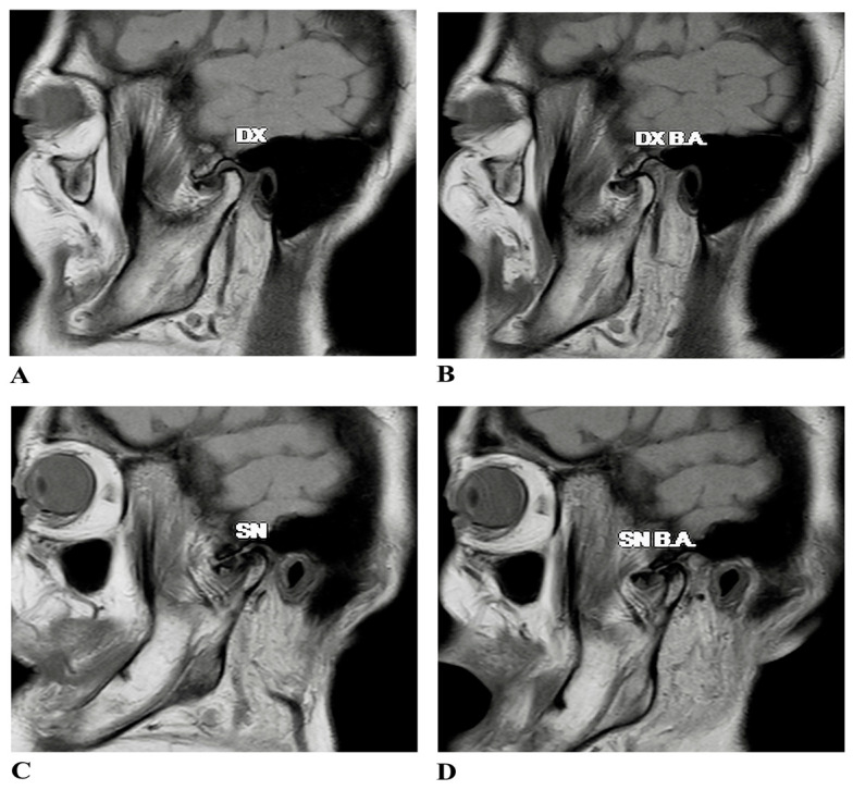
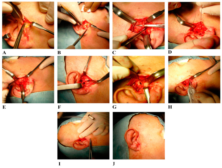
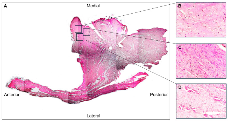
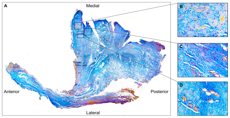
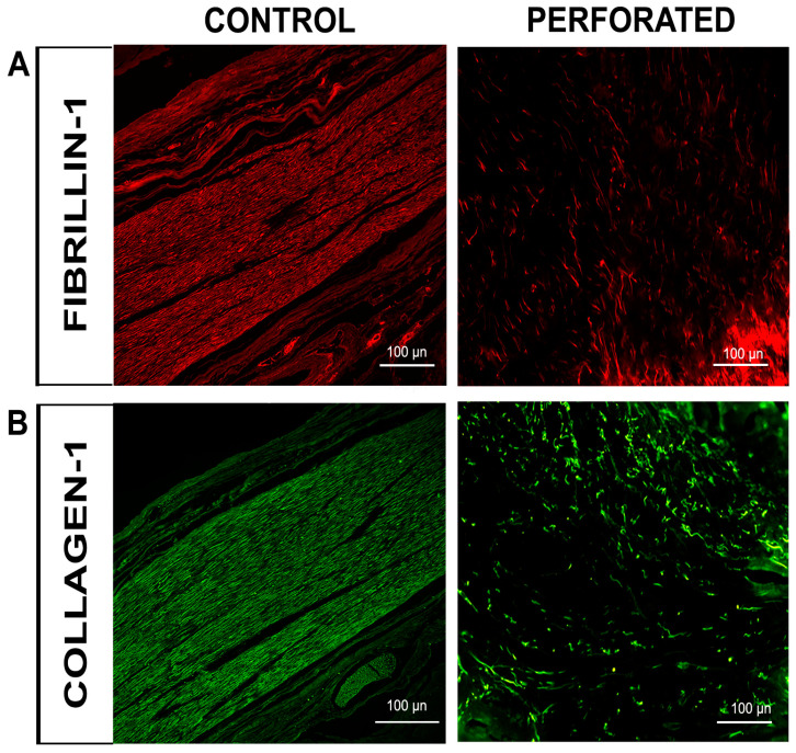
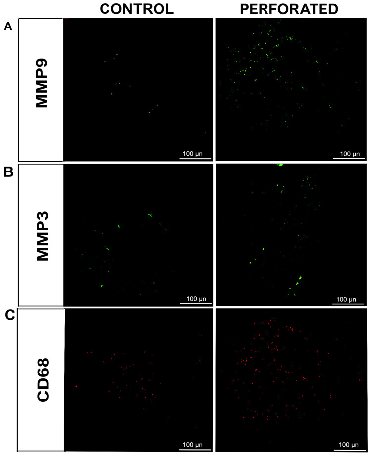
Background/Objectives: Anterior disc displacement without reduction (ADDwoR) is a temporomandibular joint (TMJ) disorder characterized by progressive dysfunction and potential complications. Persistent displacement leads to abnormal mechanical stress, predisposing the TMJ disc to structural degeneration, including perforation. This case report aimed to examine the histological and immunofluorescence characteristics of perforated disc tissue to elucidate the mechanisms contributing to its pathology. Methods: A 50-year-old patient with bilateral ADDwoR and disc perforation underwent functional arthroplasty. Tissue samples from the perforated disc were histologically analyzed using hematoxylin-eosin and Azan Mallory staining. Immunofluorescence was performed to assess the expression of collagen type I, fibrillin-1, matrix metalloproteinases (MMPs)-3 and -9, and cluster of differentiation 68 (CD68). Results: Histological analysis revealed disorganized collagen fibres and fibro-chondrocyte cell predominance in the perilesional zone, accompanied by vascular proliferation. Adjacent tissue to perforation exhibited normal fibrous organization. Immunofluorescence showed reduced collagen type I and fibrillin-1 patterns in the perilesional area, indicating an alteration in the fibrillar component of the extracellular matrix (ECM). Increased expression of MMP-3 and MMP-9, as well as elevated numbers of CD68-positive macrophages, suggested active ECM degradation and inflammation localized to the perforation site. Conclusions: This case report underscores the critical role of biomechanical stress and inflammation in disc perforation. Decreased ECM integrity, driven by altered collagen and fibrillin composition, as well as heightened MMP activity, compromises the disc's capacity to absorb and distribute mechanical loads. These findings advance our understanding of TMJ pathophysiology, emphasizing the need for therapeutic approaches that target both biomechanical stabilization and inflammation.
Keywords: ECM degeneration; anterior disc displacement; biomechanical stress; disc perforation; temporomandibular joint disorders.
Conflict of interest statement
The authors declare no conflicts of interest.
Figures
Similar articles
-
Classification of the temporomandibular joint disc displacement without reduction using MRI in the mouth-opening position.Clin Oral Investig. 2025 May 31;29(6):322. doi: 10.1007/s00784-025-06408-z. Clin Oral Investig. 2025. PMID: 40450192
-
Extracellular matrix protein composition corresponds to degenerative changes in disc displacement of the temporomandibular joint.Arch Oral Biol. 2025 Sep;177:106338. doi: 10.1016/j.archoralbio.2025.106338. Epub 2025 Jun 16. Arch Oral Biol. 2025. PMID: 40543251
-
Immunohistochemical expression of matrix metalloproteinase-9 and 13 in oral squamous cell carcinoma and their role in predicting lymph node metastasis.World J Methodol. 2025 Jun 20;15(2):94514. doi: 10.5662/wjm.v15.i2.94514. eCollection 2025 Jun 20. World J Methodol. 2025. PMID: 40548219 Free PMC article.
-
Arthroplasty versus fusion in single-level cervical degenerative disc disease.Cochrane Database Syst Rev. 2012 Sep 12;(9):CD009173. doi: 10.1002/14651858.CD009173.pub2. Cochrane Database Syst Rev. 2012. Update in: Cochrane Database Syst Rev. 2015 May 21;(5):CD009173. doi: 10.1002/14651858.CD009173.pub3. PMID: 22972137 Updated.
-
Does Temporomandibular Joint Pathology With or Without Surgical Management Affect the Stability of Counterclockwise Rotation of the Maxillomandibular Complex in Orthognathic Surgery? A Systematic Review and Meta-Analysis.J Oral Maxillofac Surg. 2017 Apr;75(4):805-821. doi: 10.1016/j.joms.2016.10.034. Epub 2016 Nov 4. J Oral Maxillofac Surg. 2017. PMID: 27889535
References
-
- Nastro E., Bonanno L., Catalfamo L., Runci M., Bramanti A., Anastasi G., Marino S., De Ponte F.S. Diffusion Tensor Imaging Reveals Morphological Alterations of the Lateral Pterygoid Muscle in Patients with Mandibular Asymmetry. Dentomaxillofac. Radiol. 2018;47:20170129. doi: 10.1259/dmfr.20170129. - DOI - PMC - PubMed
LinkOut - more resources
Full Text Sources
Miscellaneous
