

Using the Deep Learning Algorithm to Determine the Presence of Sacroiliitis from Pelvic Radiographs
- PMID: 40566530
- PMCID: PMC12193810
- DOI: 10.3390/life15060876
Using the Deep Learning Algorithm to Determine the Presence of Sacroiliitis from Pelvic Radiographs
Abstract
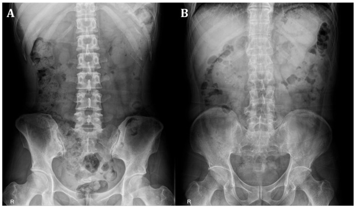
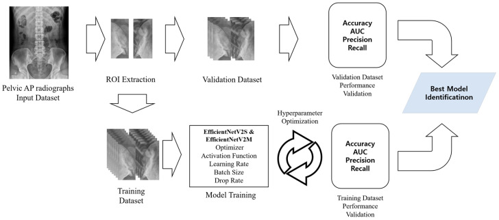
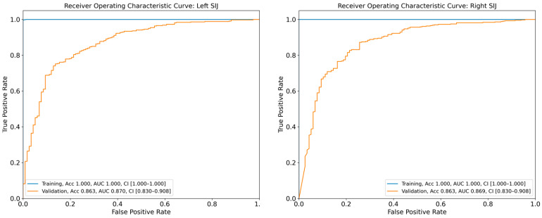
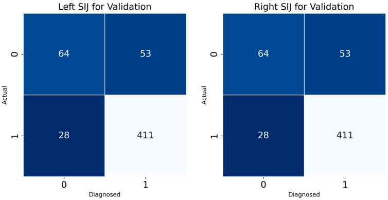
Deep learning (DL) techniques have demonstrated remarkable capabilities in recognizing complex patterns in medical imaging data. In recent years, DL has been increasingly applied in clinical medicine for disease diagnosis and progression prediction. This study aimed to develop and validate a DL model for detecting sacroiliitis using pelvic anteroposterior (AP) radiographs. We retrospectively analyzed 1853 patients with pelvic AP radiographs, including 3706 sacroiliac joints (SIJs). Pelvic AP radiographs served as input data for the DL model development, while the presence or absence of sacroiliitis confirmed by pelvic computed tomography (CT) was used as the reference standard output data. Based on CT findings, 1463 of 1853 right SIJs showed evidence of sacroiliitis, while 390 had no sacroiliitis. Similar findings were observed in the left SIJs. The dataset was split with 70% (1297 images) for training and 30% (556 images) for validation. The areas under the curve (AUC) for our DL model on the validation dataset were 0.871 (95% confidence interval (CI): 0.834-0.907) and 0.869 (95% CI: 0.834-0.907) for the left and right SIJs, respectively. Diagnostic accuracies for sacroiliitis on the left and right sides were 85.4% and 86.3%, respectively. These results demonstrate that a DL model trained on pelvic AP radiographs with CT-confirmed diagnoses can effectively aid in the diagnosis of sacroiliitis.
Keywords: computed tomography; convolutional neural network; deep learning; radiograph; sacroiliac joint; sacroiliitis.
Conflict of interest statement
The authors declare no conflicts of interest.
Figures
References
-
- Weber U., Lambert R.G., Østergaard M., Hodler J., Pedersen S.J., Maksymowych W.P. The diagnostic utility of magnetic resonance imaging in spondylarthritis: An international multicenter evaluation of one hundred eighty-seven subjects. Arthrit. Rheumat. 2010;62:3048–3058. doi: 10.1002/art.27571. - DOI - PubMed
LinkOut - more resources
Full Text Sources
