Review
. 2025 May 30;15(6):888.
doi: 10.3390/life15060888.

Voltage-Gated Ion Channels in Neuropathic Pain Signaling

Affiliations
Review

Voltage-Gated Ion Channels in Neuropathic Pain Signaling

Ricardo Felix et al. Life (Basel). .

Abstract

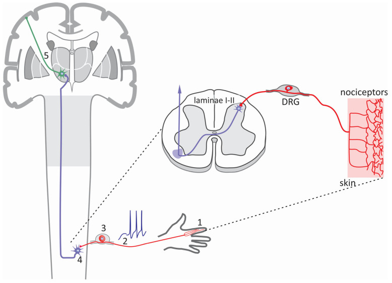
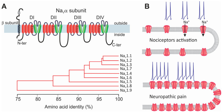
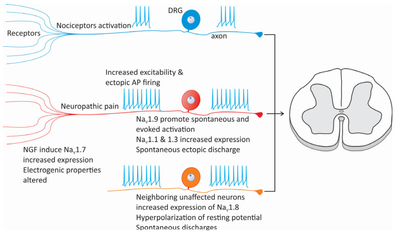
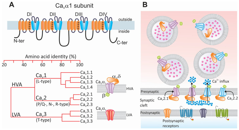
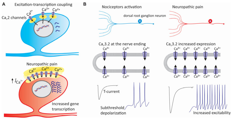
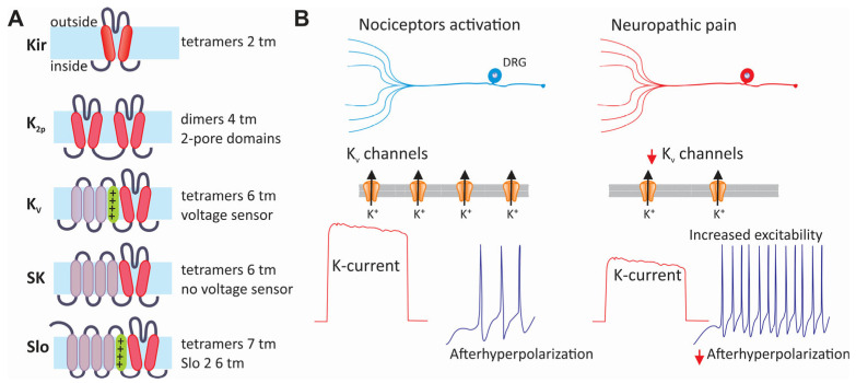
Neuropathic pain is a chronic and debilitating disorder of the somatosensory system that affects a significant proportion of the population and is characterized by abnormal responses such as hyperalgesia and allodynia. Voltage-gated ion channels, including sodium (NaV), calcium (CaV), and potassium (KV) channels, play a pivotal role in modulating neuronal excitability and pain signal transmission following nerve injury. This review intends to provide a comprehensive analysis of the molecular and cellular mechanisms by which dysregulation in the expression, localization, and function of specific NaV channel subtypes (mainly NaV1.7 and NaV1.8) and their auxiliary subunits contributes to aberrant neuronal activation, the generation of ectopic discharges, and sensitization in neuropathic pain. Likewise, special emphasis is placed on the crucial role of CaV channels, particularly CaV2.2 and the auxiliary subunit CaVα2δ, whose overexpression increases calcium influx, neurotransmitter release, and neuronal hyperexcitability, thus maintaining persistent pain states. Furthermore, KV channels (particularly KV7 channels) function as brakes on neuronal excitability, and their dysregulation facilitates the development and maintenance of neuropathic pain. Therefore, targeting specific KV channel subtypes to restore their function is also a promising therapeutic strategy for alleviating neuropathic pain symptoms. On the other hand, recent advances in the development of small molecules as selective modulators or inhibitors targeting voltage-gated ion channels are also discussed. These agents have improved efficacy and safety profiles in preclinical and clinical studies by attenuating pathophysiological channel activity and restoring neuronal function. This review seeks to contribute to guiding future research and drug development toward more effective mechanism-based treatments by discussing the molecular mechanisms underlying neuropathic pain and highlighting translational therapeutic opportunities.

Keywords: CaV channels; KV channels; NaV channels; PROTACs; calcium channels; neuropathic pain; potassium channels; sodium channels; voltage-gated ion channels.


Conflict of interest statement

The authors declare no conflicts of interest.

Figures

Figure 1
Figure 1
The sensory pathway. The somatosensory system comprises an intricate network of sensory receptors distributed throughout the skin, muscles, joints, and internal organs. These receptors include nociceptors activated in response to noxious stimuli and generate pain signals. The information generated in the periphery (1) is transmitted as action potentials (2), primarily via primary afferent fibers of the Aδ and C type that have a peripheral axon innervating the distal regions, to the DRG (3), where the soma of the sensory neurons are located. The pain signals then travel to the second-order neurons in the laminae I-II of the spinal cord (4). Finally, these signals are transmitted to third-order neurons in the thalamus (5) and then to the primary somatosensory cortex to be integrated.
Figure 2
Figure 2
Structure and classification of NaV channels and their participation in neuropathic pain. (A) The α subunit forms the ion-conducting region of NaV channels. This protein comprises four repeated homologous domains (DI–DIV), formed by six transmembrane segments connected by intracellular loops. Segment S4 (+) acts as the channel voltage sensor. Although the NaVα subunit alone can form a functional channel, it is generally associated with auxiliary subunits β (blue; NaVβ1–NaVβ4) that regulate its biophysical properties, its trafficking to the membrane, and its interaction with proteins extrinsic to the channel (upper panel). Nine α subunits have been identified (NaV1.1α–NaV1.9α) that share a similar membrane topology, each encoded by a different gene with different properties. The phylogenetic tree illustrates the amino acid sequence similarity of the mammal NaVα subunits encoding the nine identified NaV channels (lower panel). (B) In neuropathic pain, significant alterations occur in both the functional expression and the activity of NaV channels. These changes affect neuronal excitability, which translates as increased sensitivity to pain and produces hyperalgesia and allodynia.
Figure 3
Figure 3
Changes in NaV channel expression and cellular excitability in neuropathic pain. Alterations in the expression of various subtypes of sodium channels, such as NaV1.1, NaV1.3, NaV1.7, NaV1.8, and NaV1.9, can increase cellular excitability, reducing the activation threshold of nociceptors. Similarly, after nerve injury, neurons adjacent to the damaged area may experience changes in the expression of NaV channels, particularly NaV1.3 and NaV1.8, which causes the development of ectopic foci of neuronal activity. On the other hand, the expression of NaV1.8 channels in neurons neighboring an injured nerve can also be compromised, contributing to the maintenance of neuropathic pain.
Figure 4
Figure 4
Structure and classification of CaV channels and their participation in neurotransmission. (A) Schematic representation of the CaVα1 subunit illustrating its membrane topology. Like NaV channels and some members of the KV channel family, the main CaVα1 subunit is a protein composed of four relatively conserved homologous repeat domains (DI–DIV) containing six α helices each (upper panel). The fourth α helix of each repeat domain contains a sequence of regularly spaced positively charged (+) basic residues that sense changes in transmembrane voltage. The loops connecting the repeat domains, as well as the amino and carboxyl termini, are intracellular. The lower left panel shows a phylogenetic tree illustrating the evolutionary relationship among members of the CaV channel family. The structural homology comparison is based on the alignment of the human channels. LVA and HVA stand for high- and low-voltage-activated, respectively. HVA channels (CaV1 and CaV2) are oligomeric complexes whose composition, in addition to the pore-forming CaVα1 subunit, includes two auxiliary subunits called CaVβ and CaVα2δ (shown in green and orange, respectively). On the other hand, LVA (CaV3) channels function as monomers of the main CaVα1 subunit (lower right panel). (B) In response to membrane depolarization caused by the arrival of an AP, presynaptic CaV channels allow for the entry of calcium ions (blue dots) from the extracellular space to the synaptic terminals. The most relevant channel subtypes involved in this event are CaV2.1 and CaV2.2 (shown in purple and blue, respectively). Increased intracellular calcium concentration at active sites promotes fusion of neurotransmitter-containing vesicles through the SNARE complex. Neurotransmitters (pink dots) released into the synaptic cleft diffuse until they bind to their receptors located on the postsynaptic membrane.
Figure 5
Figure 5
Contribution of CaV channels to the pathogenesis of neuropathic pain. (A) In addition to its effects on the release of neurotransmitters, alteration in the expression of CaV2 channels participates in the pathophysiology of neuropathic pain by affecting the excitation–transcription coupling. This fundamental cell process links electrical activity in excitable cells to gene transcription. This implies that the calcium, once inside the cells, can activate transcription factors, either directly or through protein kinases and second messengers that control the activity of these factors. (B) Overexpression of CaV3 channels in sensory neurons during neuropathic pain increases their excitability, decreases the firing threshold of afferent fibers, and favors repetitive firing.
Figure 6
Figure 6
Classification and contribution of KV channels to neuropathic pain. (A) The family of potassium channels arranged according to the structure of their main α subunits. The members of this family can be grouped into those formed by tetramers of two (Kir) or dimers of four (two pores) transmembrane segments. Likewise, channels formed by six transmembrane segments, the predominant voltage-sensitive potassium channels, assemble into a tetramer to form a functional channel. The same is also valid for the small-conductance calcium-activated potassium (SK) channels and the large-conductance channels activated by both changes in intracellular calcium and membrane voltage. (B) The expression of KV channels is often decreased in neuropathic pain. This decreases the outward current, which usually helps to stabilize the membrane potential and opposes excitatory signals. The decreased activity of KV channels during neuropathic pain may cause an increase in neuronal excitability, affecting the frequency and duration of APs.

Similar articles

Cited by

References

    1. Finnerup N.B., Kuner R., Jensen T.S. Neuropathic pain: From mechanisms to treatment. Physiol. Rev. 2021;101:259–301. doi: 10.1152/physrev.00045.2019. - DOI - PubMed
    1. van Hecke O., Austin S.K., Khan R.A., Smith B.H., Torrance N. Neuropathic pain in the general population: A systematic review of epidemiological studies. Pain. 2014;155:654–662. doi: 10.1016/j.pain.2013.11.013. - DOI - PubMed
    1. Colloca L., Ludman T., Bouhassira D., Baron R., Dickenson A.H., Yarnitsky D., Freeman R., Truini A., Attal N., Finnerup N.B., et al. Neuropathic pain. Nat. Rev. Dis. Primers. 2017;3:17002. doi: 10.1038/nrdp.2017.2. - DOI - PMC - PubMed
    1. St John Smith E. Advances in understanding nociception and neuropathic pain. J. Neurol. 2018;265:231–238. doi: 10.1007/s00415-017-8641-6. - DOI - PMC - PubMed
    1. Bagal S.K., Marron B.E., Owen R.M., Storer R.I., Swain N.A. Voltage-gated sodium channels as drug discovery targets. Channels. 2015;9:360–366. doi: 10.1080/19336950.2015.1079674. - DOI - PMC - PubMed

LinkOut - more resources