. 2025 Jun 5;15(6):918.
doi: 10.3390/life15060918.

Trophoblast Extracellular Vesicles as Modulators of Keratinocyte Stress Response and Senescence

Affiliations

Trophoblast Extracellular Vesicles as Modulators of Keratinocyte Stress Response and Senescence

Mirjana Nacka-Aleksić et al. Life (Basel). .

Abstract

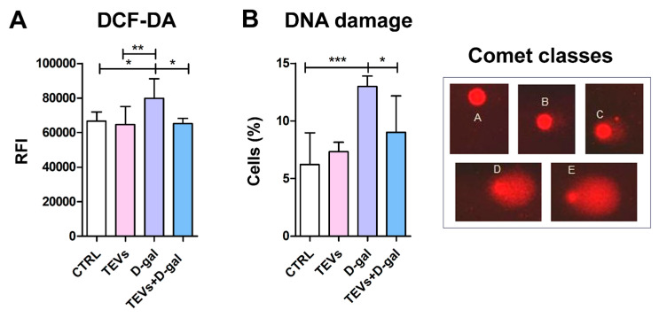
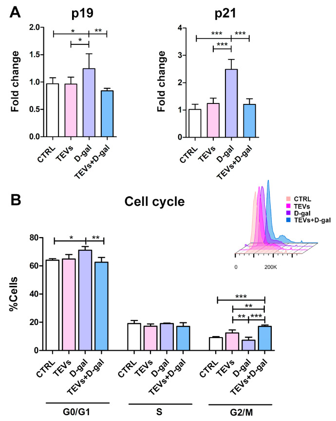
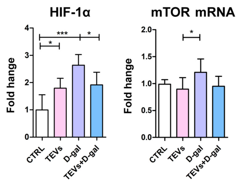
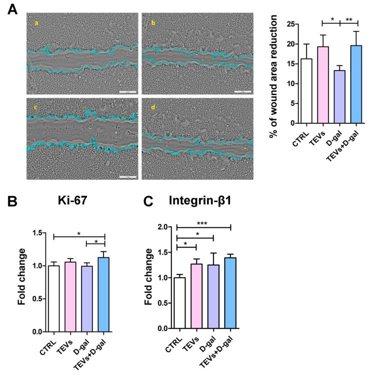
Keratinocyte stress, caused by various intrinsic and extrinsic factors, contributes to the overall aging process. D-galactose-induced metabolic/oxidative stress is a commonly used in vitro model for studying premature aging. Due to their rich composition of bioactive molecules that influence critical pathways in cellular aging and rejuvenation, placental derivatives have a well-established history in anti-aging skincare and therapy. However, trophoblast-derived extracellular vesicle (TEV) effects on D-galactose-induced premature aging in keratinocytes have not been investigated yet. TEV pretreatment for 24 h enhanced cellular resilience against D-galactose-induced stress, judging by the downregulated expression of senescence- and stress-associated markers (p19 and p21, HIF-1α, mTOR), and reduced production of reactive oxygen species and DNA damage. Additionally, TEV pretreatment enhanced keratinocyte proliferation and integrin-β1 subunit expression upon D-galactose exposure, most likely contributing to more efficient wound closure. In conclusion, this study underscores the potential of TEVs to modify expression of stress- and senescence-related proteins in keratinocytes and improve their wound healing properties. Their regenerative and protective characteristics position TEVs as promising candidates for developing innovative procedures to address skin conditions related to premature aging.

Keywords: aging; placenta; regenerative medicine; skin; wound healing.


Conflict of interest statement

The authors declare no conflicts of interest.

Figures

Figure 1
Figure 1
Characterization of trophoblast-derived extracellular vesicles (TEVs) by nanoparticle tracking analysis (NTA), transmission electron microscopy (TEM), and super-resolution dSTORM imaging. (A) Representative frame from ZetaView Quatt PMX-430 showing concentration and size measurement of TEVs by NTA. (B) Representative TEM native image of TEVs (inset displays zoomed-in vesicle). (C) Representative ONI super-resolution nanoimaging TEV captures. Antibodies targeted against CD9-AlexaFluor™ 488 (cyan), CD63-AlexaFluor™ 561 (yellow), and CD81-AlexaFluor™ 647 (purple) allowed for detection of their surface expression of immobilized TEVs (see Materials and Methods).
Figure 2
Figure 2
Trophoblast-derived extracellular vesicle (TEV) pretreatment diminishes reactive oxygen species (ROS) level and DNA damage in keratinocytes exposed to D-gal. Bar graphs indicate (A) relative fluorescence intensity (RFI) of dihydrofluorescein dye (DCF-DA) in keratinocytes, which correlates with intracellular ROS levels, and (B) percentage of cells with DNA damage, assessed by the alkaline comet assay. Representative microscopic images (40× magnification) show keratinocyte nuclei with damaged DNA (comets). Visual scoring of the comets to five classes according to the amount of fragmented DNA in the comet tail: A. no damage, <5%; B. low-level damage, 5–20%; C. medium-level damage, 20–40%; D. high-level damage, 40–75%; E. total damage, >75%. Cumulative data from two experiments (3–4 replicates) are expressed as mean + SD. * p < 0.05; ** p < 0.01; *** p < 0.001.
Figure 3
Figure 3
Pretreatment of keratinocytes with trophoblast-derived extracellular vesicles (TEVs) prevents upregulation of senescence markers and cell cycle arrest. Bar graphs indications: (A) Expression of cyclin-dependent kinase inhibitors p19 and p21 in keratinocytes, as determined by cELISA. Cumulative data from two experiments (3 replicates) are shown as mean fold change relative to the unexposed cells (CTRL) + SD. (B) Representative overlaid flow cytometry histograms indicate the frequency of cells in G0/G1, S, G2/M phases of the cell cycle. Data (mean + SD) are representative from one of two experiments (4 replicates) with consistent results. * p < 0.05; ** p < 0.01; *** p < 0.001.
Figure 4
Figure 4
Trophoblast-derived extracellular vesicle (TEV) pretreatment ameliorates HIF-1α and mTOR upregulation in D-gal-exposed keratinocytes. Bar graphs indicate protein expression of HIF-1α and mRNA expression of mTOR in keratinocytes, as determined by cELISA and qPCR, respectively. Cumulative data from two experiments (3 replicates) are shown as mean fold change relative to the unexposed cells (CTRL) + SD. * p < 0.05; *** p < 0.001.
Figure 5
Figure 5
Trophoblast-derived extracellular vesicle (TEV) pretreatment enhances keratinocyte migration and Ki-67 and integrin-β1 subunit expression after D-gal exposure. (A) Representative phase-contrast photomicrographs ((a) CTRL; (b) TEVs; (c) D-gal; (d) TEVs+D-gal) show keratinocyte migration after 24 h. Bar graphs indicate (A) the percentage of wound closure relative to the initial scratch area and the protein level of (B) Ki-67 and (C) integrin-β1 subunit expression in keratinocytes, as determined by cELISA. Cumulative data from two experiments (3–5 replicates) are shown as mean fold change relative to the unexposed cells (CTRL) + SD. * p < 0.05; ** p < 0.01; *** p < 0.001.

Similar articles

References

    1. Kanitakis J. Anatomy, Histology and Immunohistochemistry of Normal Human Skin. Eur. J. Dermatol. 2002;12:390–399; quiz 400–401. - PubMed
    1. Wong Q.Y.A., Chew F.T. Defining Skin Aging and Its Risk Factors: A Systematic Review and Meta-Analysis. Sci. Rep. 2021;11:22075. doi: 10.1038/s41598-021-01573-z. - DOI - PMC - PubMed
    1. Kuang L., Li C. ΔNp63α-Mediated Epigenetic Regulation in Keratinocyte Senescence. Epigenetics. 2023;18:2173931. doi: 10.1080/15592294.2023.2173931. - DOI - PMC - PubMed
    1. Nguyen H.P., Katta R. Sugar Sag: Glycation and the Role of Diet in Aging Skin. Ski. Ther. Lett. 2015;20:1–5. - PubMed
    1. Danby F.W. Nutrition and Aging Skin: Sugar and Glycation. Clin. Dermatol. 2010;28:409–411. doi: 10.1016/j.clindermatol.2010.03.018. - DOI - PubMed

LinkOut - more resources