

The Platelet-to-Lymphocyte Ratio (PLR) as a Non-Invasive Biomarker for Cervical Malignancy in Conization Patients
- PMID: 40566623
- PMCID: PMC12194268
- DOI: 10.3390/life15060971
The Platelet-to-Lymphocyte Ratio (PLR) as a Non-Invasive Biomarker for Cervical Malignancy in Conization Patients
Abstract
Background: Cervical cancer continues to pose a significant global health challenge, particularly in low-resource regions with limited access to advanced diagnostics. Cervical conization can occasionally uncover invasive carcinoma in patients initially suspected of having only pre-invasive lesions. This study assessed the platelet-to-lymphocyte ratio (PLR) as a potential predictive biomarker for identifying invasive disease in patients undergoing a loop electrosurgical excision procedure (LEEP).
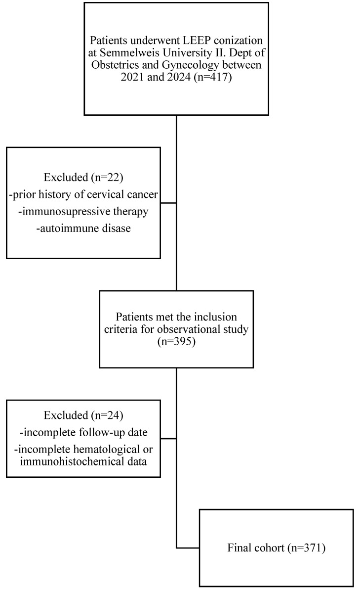
Methods: A retrospective study was conducted on 371 patients who underwent LEEP conization for cervical dysplasia. Preoperative PLR values were collected and compared across final histopathological categories (negative, low-grade, high-grade, invasive carcinoma) using the Kruskal-Wallis test, followed by Mann-Whitney U tests for pairwise comparisons. Receiver operating characteristic (ROC) analysis was used to evaluate diagnostic accuracy.
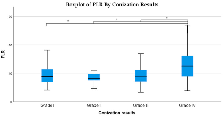
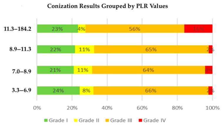
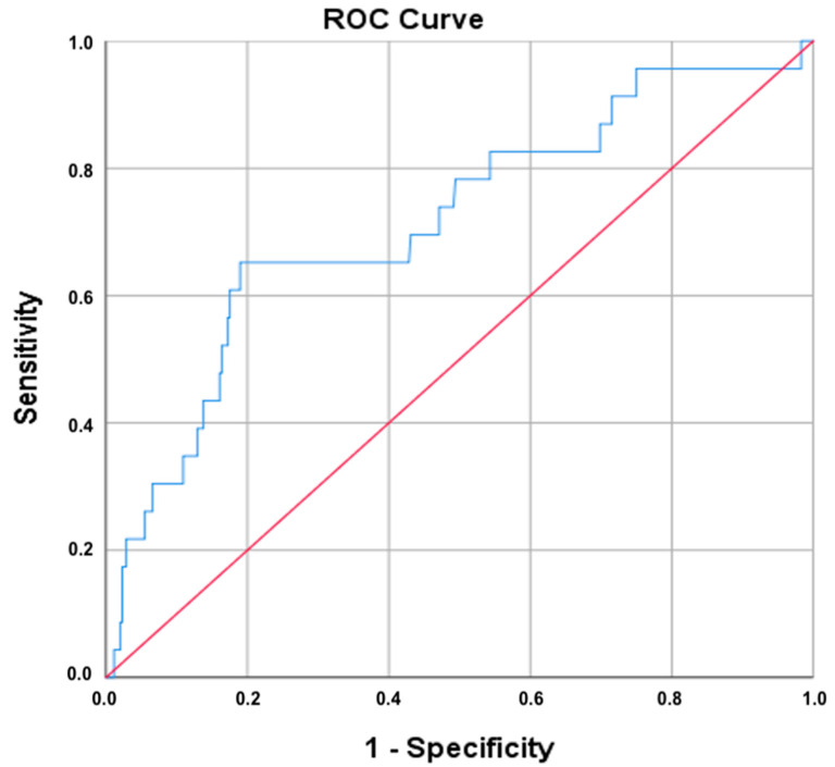
Results: PLR values above 7.7 were significantly associated with HPV positivity, increasing with histopathological severity. There were significant PLR differences across the outcome groups (p = 0.005), with notably higher values in cases of invasive carcinoma (p < 0.01). ROC analysis showed moderate diagnostic utility (AUC ≈ 0.72); at a PLR cutoff of ~11.9, sensitivity was 65% and specificity 81%.
Conclusions: The PLR cutoff of 7.7 was associated with HPV positivity, while a higher cutoff of 11.93 was identified for predicting invasive cervical cancer. These findings support that preoperative PLR is a non-invasive, clinically relevant marker correlated with lesion severity, offering potential for preoperative risk stratification, particularly where advanced diagnostics are limited.
Keywords: HPV DNA; ROC analysis; biomarkers; cervical cancer; invasive carcinoma; platelet-to-lymphocyte ratio (PLR); risk stratification; systemic inflammation.
Conflict of interest statement
The authors declare no conflicts of interest.
Figures

Similar articles
-
Signs and symptoms to determine if a patient presenting in primary care or hospital outpatient settings has COVID-19.Cochrane Database Syst Rev. 2022 May 20;5(5):CD013665. doi: 10.1002/14651858.CD013665.pub3. Cochrane Database Syst Rev. 2022. PMID: 35593186 Free PMC article.
-
Medical and surgical interventions for the treatment of usual-type vulval intraepithelial neoplasia.Cochrane Database Syst Rev. 2016 Jan 5;2016(1):CD011837. doi: 10.1002/14651858.CD011837.pub2. Cochrane Database Syst Rev. 2016. PMID: 26728940 Free PMC article.
-
Non-invasive diagnostic tests for Helicobacter pylori infection.Cochrane Database Syst Rev. 2018 Mar 15;3(3):CD012080. doi: 10.1002/14651858.CD012080.pub2. Cochrane Database Syst Rev. 2018. PMID: 29543326 Free PMC article.
-
Intraoperative frozen section analysis for the diagnosis of early stage ovarian cancer in suspicious pelvic masses.Cochrane Database Syst Rev. 2016 Mar 1;3(3):CD010360. doi: 10.1002/14651858.CD010360.pub2. Cochrane Database Syst Rev. 2016. PMID: 26930463 Free PMC article.
-
Interventions targeted at women to encourage the uptake of cervical screening.Cochrane Database Syst Rev. 2021 Sep 6;9(9):CD002834. doi: 10.1002/14651858.CD002834.pub3. Cochrane Database Syst Rev. 2021. PMID: 34694000 Free PMC article.
References
LinkOut - more resources
Full Text Sources
