Review
. 2025 Jun 25;6(7):e70258.
doi: 10.1002/mco2.70258. eCollection 2025 Jul.

Advancing Design Strategy of PROTACs for Cancer Therapy

Affiliations
Review

Advancing Design Strategy of PROTACs for Cancer Therapy

Hang Luo et al. MedComm (2020). .

Abstract

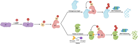
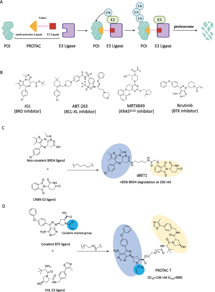
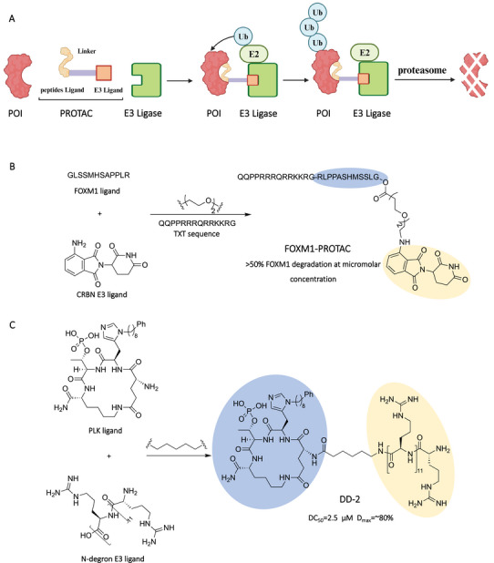
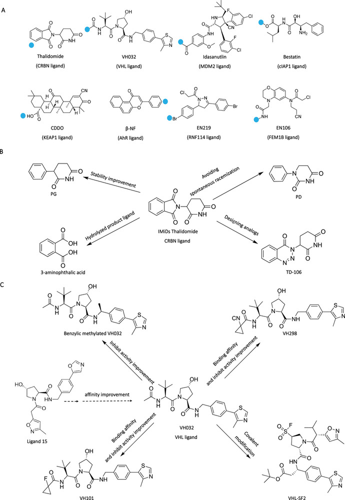
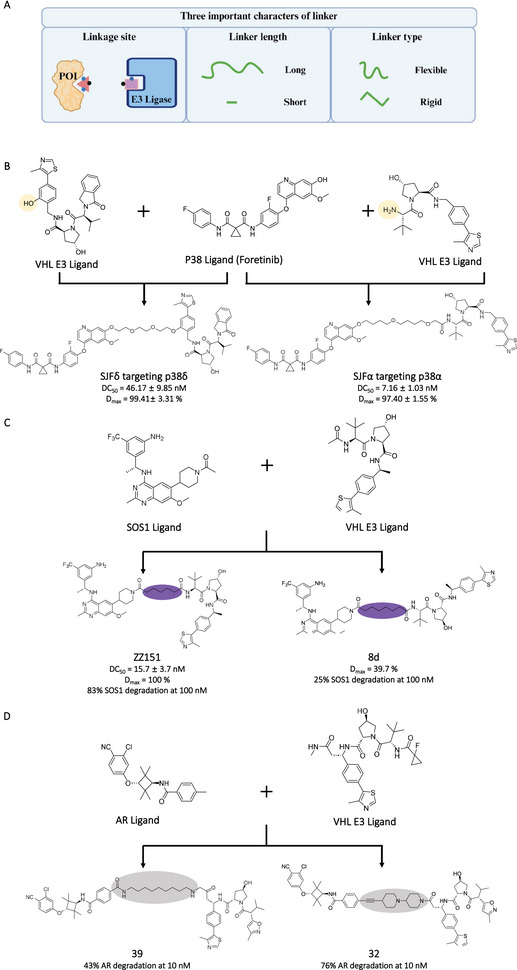
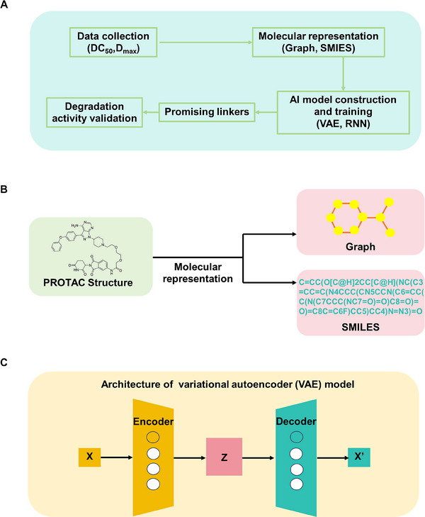
Proteolysis targeting chimeras (PROTACs) have emerged as a groundbreaking class of anticancer therapeutics. These bifunctional molecules harness the endogenous ubiquitin-proteasome system to facilitate the degradation of targeted proteins of interest (POIs). Notably, the clinical translation of PROTACs has gained substantial momentum, with many PROTAC candidates targeting various cancers currently undergoing clinical trials (Phase I-III). However, the rational design of high-efficacy PROTAC compounds remains a significant challenge. In this review, we presented a comprehensive overview of POI ligands, E3 ligands, and their interconnected linkers in PROTAC design, including their generation, structural optimization, and contribution to degradation efficiency and selectivity. Particularly, we analyzed the distinct preferences of various types of POI ligands (small molecule, nucleic acid, and peptide) toward specific targets. Furthermore, we emphasized the significant role of artificial intelligence technology in PROTAC design, including POI/E3 ligands discovery and linkers generation or optimization. We also summarized the applications and challenges of PROTACs in cancer therapy. Finally, we discussed the future development of PROTAC by combining multidisciplinary technologies and novel modalities for cancer therapy. Overall, this review aims to provide valuable insights for advancing PROTAC design strategies for cancer therapy.

Keywords: E3 ligand; POI ligand; artificial intelligence; cancer therapy; linker design; proteolysis targeting chimeras (PROTACs).


Conflict of interest statement

All authors declare no conflicts of interest.

Figures

FIGURE 1
FIGURE 1
Mechanism of action of UPS system‐mediated endogenous protein degradation and PROTACs‐mediated targeted protein degradation. First, the E1 could activate ubiquitin in an ATP‐dependent manner. Subsequently, the ubiquitin could be transferred on E2, and then the E3–E2–ubiquitin complex could be formed. For the endogenous protein ubiquitination, the polyubiquitin could be labeled on the endogenous protein by E3 ligase. For the POI targeted ubiquitination, the polyubiquitin could be formed on the POI by E3 ligase catalysis after forming a POI/PROTAC/E3 ternary complex. Finally, the ubiquitinated endogenous protein or POI could be transported to proteasome for degradation. ATP: adenosine triphosphate. The Figure 1 was created in BioRender.com.
FIGURE 2
FIGURE 2
Small molecules as POI ligands in PROTACs. (A) The action mode of small molecules‐based PROTACs. (B) The chemical structures of representative small molecule POI ligands including JQ1, ABT‐263, MRTX849, and ibrutinib. (C) Design of noncovalent small molecule‐based PROTAC (dBET1) by connecting the noncovalent BRD4 ligand and a CRBN E3 ligand. (D) Design of covalent small molecule‐based PROTAC (PROTAC 7) by connecting the covalent BTK ligand and a VHL E3 ligand. The blue part represents the covalent reaction group, and the light blue part and light orange part represent POI ligand and E3 ligand, respectively. The Figure 2A was created in BioRender.com.
FIGURE 3
FIGURE 3
Nucleic acids as POI ligands in PROTACs. (A) The action mode of nucleic acid‐based PROTACs. (B) Design of single strand RNA‐based PROTAC (ORN3P1) by connecting the single strand RNA targeting Lin28A and a VHL E3 ligand. (C) Design of double strand DNA‐based PROTAC (OP‐V1) by connecting the double strand DNA targeting LEF1 and a VHL E3 ligand. (D) Design of aptamer‐based PROTAC (dNCL#T1) by connecting the aptamer targeting NCL and a CRBN ligand. The light blue part and light orange part represent POI ligand and E3 ligand, respectively. The Figure 3A was created in BioRender.com.
FIGURE 4
FIGURE 4
Peptides as POI ligands in PROTACs. (A) The action mode of peptides‐based PROTACs. (B) Design of peptide sequence‐based PROTAC (FOXM1–PROTAC) by connecting the peptide sequence targeting FOXM1 and a CRBN ligand. (C) Design of the cyclic peptide‐based PROTAC (DD‐2) by connecting the cyclic peptide targeting PLK and a N‐dergon E3 ligand. The blue light part and light orange part represent POI ligand and E3 ligand, respectively. The Figure 4A was created in BioRender.com.
FIGURE 5
FIGURE 5
E3 ligands in PROTAC design. (A) Representative E3 ligands in PROTACs, where the blue dot represents the attachment sites. (B) Design and optimization of CRBN E3 ligand Thalidomide. (C) Design and optimization of VHL E3 ligand VH032.
FIGURE 6
FIGURE 6
The linkers in PROTAC design. (A) Three important characters of linker in PROTAC design. (B) Different linker attachment sites in E3 ligands in PROTACs (SJFδ and SIFα), where the orange parts represent linker attachment sites. (C) Different linker length in PROTACs (ZZ51 and 8d), where the purple parts represent linkers. (D) Different linker composition in PROTACs (39 and 32), where the gray parts represent linkers. The Figure 6A was created in BioRender.com.
FIGURE 7
FIGURE 7
AI‐based linkers in PROTAC design. (A) The flow chart of AI‐based linker design strategies, including the data collection, molecular representation, model construction and training, promising linker output, and degradation efficiency validation. (B) Molecular representations (graph and SMILES) used in AI models. (C) Architectures of VAE model.

Similar articles

References

    1. Sakamoto K. M., Kim K. B., Kumagai A., Mercurio F., Crews C. M., and Deshaies R. J., “Protacs: Chimeric Molecules That Target Proteins to the Skp1‐Cullin‐F Box Complex for Ubiquitination and Degradation,” PNAS 98, no. 15 (2001): 8554–8559. - PMC - PubMed
    1. Gadd M. S., Testa A., Lucas X., et al., “Structural Basis of PROTAC Cooperative Recognition for Selective Protein Degradation,” Nature Chemical Biology 13, no. 5 (2017): 514–521. - PMC - PubMed
    1. Glickman M. H. and Ciechanover A., “The Ubiquitin‐proteasome Proteolytic Pathway: Destruction for the Sake of Construction,” Physiological Reviews 82, no. 2 (2002): 373–428. - PubMed
    1. Ma B., Fan Y., Zhang D., et al., “De Novo Design of an Androgen Receptor DNA Binding Domain‐targeted Peptide PROTAC for Prostate Cancer Therapy,” Advancement of Science 9, no. 28 (2022): 2201859. - PMC - PubMed
    1. Lee S., Kim H. R., Woo Y., et al., “UBX‐390: A Novel Androgen Receptor Degrader for Therapeutic Intervention in Prostate Cancer,” Advancement of Science 11, no. 33 (2024): 2400398. - PMC - PubMed

LinkOut - more resources