. 2021 Jul 28;1(3):100034.
doi: 10.1016/j.ynirp.2021.100034. eCollection 2021 Sep.

Basal ganglia activation localized in MEG using a reward task

Affiliations

Basal ganglia activation localized in MEG using a reward task

Linnea Sepe-Forrest et al. Neuroimage Rep. .

Abstract

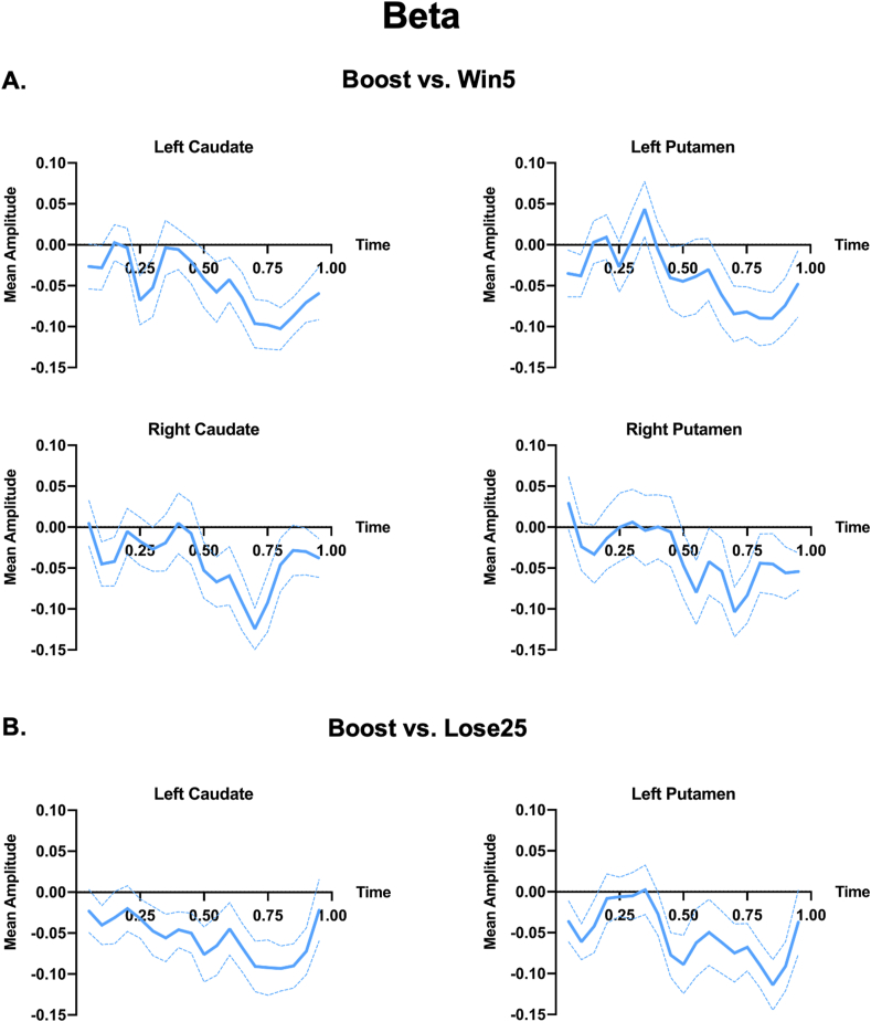
The basal ganglia are a crucial component of neural networks underlying reward response and many other behaviors. Magnetoencephalography (MEG) can be used to non-invasively study the spatiotemporal dynamics of activity in neural networks. However, challenges associated with detecting deep sources has caused many to doubt the ability of MEG to detect basal ganglia signals. In this study, we employed a gambling task to assess the feasibility of using MEG to investigate basal ganglia-cortical networks during reward processing. Participants gambled to win or lose 5 or 25 cents and received unexpected high-value rewards of 50 cents at random intervals. We contrasted activity between reward conditions in the beta (15-30 Hz), gamma (30-60 Hz), and high gamma (60-150 Hz) bands. We found differences in oscillatory power in the beta and high gamma bands while contrasting the large reward condition with both the small reward and large loss conditions. Basal ganglia activity was localized to the caudate, putamen, and globus pallidum, while cortical activity appeared primarily in parietal and temporal areas. Our results show robust basal ganglia power differences in response to reward and corroborate animal literature showing beta and high gamma activation in the striatum. This experiment demonstrates that it is possible to study basal ganglia activity using MEG and reveals specific characteristics of the normal reward response that will inform future research.

Keywords: Basal ganglia; Cortico-striatal; Gambling; Magnetoencephalography; Reward.


Conflict of interest statement

The authors declare that they have no known competing financial interests or personal relationships that could have appeared to influence the work reported in this paper.

Figures

Fig. 1
Fig. 1
Experimental design. A) Choice, Loss, Win, and Outcome trials. During the choice period, participants, 25 and 5. Within this period, participants ‘bet’ on a number by pressing the corresponding button (right button for the right number, left button for the left number). During the outcome period in trials without a Boost outcome, the box surrounding the chosen number would change colors with green indicating a win and red indicating a loss of the number of cents the subject chose to bet on. In trials with a Boost outcome, the box would turn yellow and the subject would gain 50 cents regardless of the chosen number. B). Timing information for each period within a single trial. The 1 s interstimulus trial consisted of fixation mark under the running total of monetary wins. Following the interstimulus trial was a 2 s choice period and 1 s outcome period.
Fig. 2
Fig. 2
Boost vs. Win5 subcortical and cortical activation differences between the two conditions thresholded at p = .001. Cortical data was displayed using SUMA-AFNI surface mapper (Saad and Reynolds, 2012). The activation color scale is the Cohen's d effect size of the difference. Max ES signifies the maximum effect size in each analysis, which was used to set the maximum and minimum of the color scale for each plot. Blue indicates greater activation in the Win5 condition, while red indicates greater activation in the Boost condition. A) Beta (15–30 Hz) band activation during the 0.25-0.75s and 0.5-1s time windows. B) High gamma (60–150 Hz) band activation during the 0–.5s, 0.25-0.75s, and 0.5-1s time windows.
Fig. 3
Fig. 3
Boost vs. Lose25 subcortical and cortical activation differences between the two conditions thresholded at p = .001. The activation color scale is the Cohen's d effect size of the difference. Max ES signifies the maximum effect size in each analysis, which was used to set the maximum and minimum of the color scale for each plot. Blue indicates greater activation in the Lose25 condition, while red indicates greater activation in the Boost condition. A) Beta (15–30 Hz) band activation during the 0.25-0.75s and 0.5-1s time windows. B) High gamma (60–150 Hz) band activation during the 0.25-0.75s, and 0.5-1s time windows.
Fig. 4
Fig. 4
Fractional change in log beta power in basal ganglia regions throughout the 1 s reward outcome period. Time series analysis shows mean amplitude over .1s intervals, plotted at the center of the interval. A) Boost vs. Win5 activation differences in left caudate, left putamen, right caudate, and right putamen. B) Boost vs. Lose25 activation differences in left caudate and left putamen.
Fig. 5
Fig. 5
Fractional change in log high gamma power in basal ganglia regions throughout the 1 s reward outcome period. Time series analysis shows mean amplitude over .1s intervals, plotted at the center of the interval. Boost vs. Win5 activation differences shown in left caudate, right caudate, right putamen, and right globus pallidum.

References

    1. Aarts E., Helmich R.C., Janssen M.J., Oyen W.J., Bloem B.R., Cools R. Aberrant reward processing in Parkinson's disease is associated with dopamine cell loss. Neuroimage. 2012;59(4):3339–3346. doi: 10.1016/j.neuroimage.2011.11.073. - DOI - PubMed
    1. Amemori K.I., Amemori S., Gibson D.J., Graybiel A.M. Striatal beta oscillation and neuronal activity in the primate caudate nucleus differentially represent valence and arousal under approach-avoidance conflict. Front. Neurosci. 2020;14:89. doi: 10.3389/fnins.2020.00089. - DOI - PMC - PubMed
    1. Ahsan R.L., Allom R., Gousias I.S., Habib H., Turkheimer F.E., Free S., Hammers A. Volumes, spatial extents and a probabilistic atlas of the human basal ganglia and thalamus. Neuroimage. 2007;38(2):261–270. doi: 10.1016/j.neuroimage.2007.06.004. - DOI - PubMed
    1. Arsalidou M., Vijayarajah S., Sharaev M. Basal ganglia lateralization in different types of reward. Brain Imaging Behav. 2020 doi: 10.1007/s11682-019-00215-3. - DOI - PubMed
    1. Asaad W.F., Eskandar E.N. Encoding of both positive and negative reward prediction errors by neurons of the primate lateral prefrontal cortex and caudate nucleus. J. Neurosci. 2011;31(49):17772–17787. doi: 10.1523/JNEUROSCI.3793-11.2011. - DOI - PMC - PubMed

LinkOut - more resources