. 2025;7(6):967-978.
doi: 10.1038/s42256-025-01040-8. Epub 2025 Jun 18.

Next-generation phenotyping of inherited retinal diseases from multimodal imaging with Eye2Gene

Affiliations

Next-generation phenotyping of inherited retinal diseases from multimodal imaging with Eye2Gene

Nikolas Pontikos et al. Nat Mach Intell. 2025.

Abstract

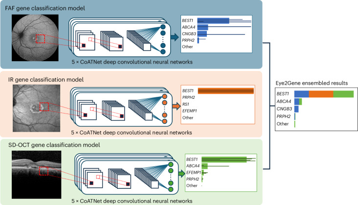
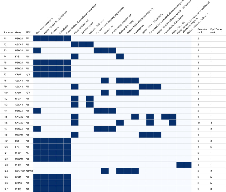
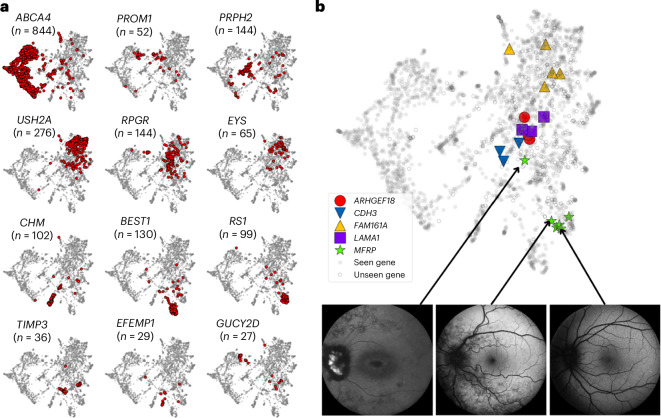
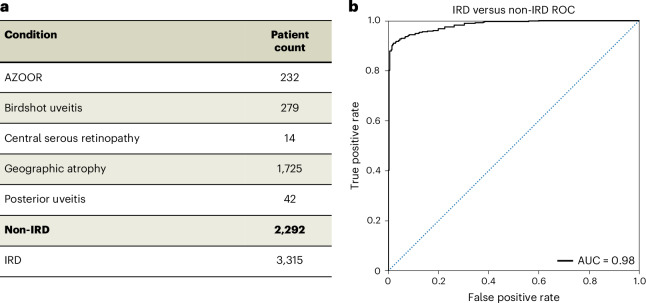
Rare eye diseases such as inherited retinal diseases (IRDs) are challenging to diagnose genetically. IRDs are typically monogenic disorders and represent a leading cause of blindness in children and working-age adults worldwide. A growing number are now being targeted in clinical trials, with approved treatments increasingly available. However, access requires a genetic diagnosis to be established sufficiently early. Critically, the timely identification of a genetic cause remains challenging. We demonstrate that a deep learning algorithm, Eye2Gene, trained on a large multimodal imaging dataset of individuals with IRDs (n = 2,451) and externally validated on data provided by five different clinical centres, provides better-than-expert-level top-five accuracy of 83.9% for supporting genetic diagnosis for the 63 most common genetic causes. We demonstrate that Eye2Gene's next-generation phenotyping can increase diagnostic yield by improving screening for IRDs, phenotype-driven variant prioritization and automatic similarity matching in phenotypic space to identify new genes. Eye2Gene is accessible online (app.eye2gene.com) for research purposes.

Keywords: Genetic testing; Image processing; Molecular medicine; Retinal diseases.


Conflict of interest statement

Competing interestsN.P., W.W., M.M. and A.R.W. are patent holders of PCT/EP2023/076614 filed by UCL Business. I.M. is a cofounder, share-holder and director of Phenopolis Ltd, the software company that developed www.eye2gene.com. N.P. is a cofounder and former share-holder and director of Phenopolis Ltd. A.Y.L. reports grants from Santen, personal fees from Genentech, personal fees from US FDA, personal fees from Johnson and Johnson, grants from Carl Zeiss Meditec, personal fees from Gyroscope, non-financial support from Microsoft and grants from Regeneron, outside the submitted work. M.M. has received consultancy or advisory board fees from MeiraGTx, Janssen Pharmaceuticals, Saliogen and Octant; travel grants from MeiraGTx and Janssen Pharmaceuticals and stock options from MeiraGTx. The other authors declare no competing interests.

Figures

Fig. 1
Fig. 1. Eye2Gene model.
Eye2Gene provides IRD-gene prediction given a retinal scan of one of three imaging modalities (FAF, IR or SD-OCT) for up to 63 gene classes. Images are initially resized to 256 by 256 pixels and rescaled to the range [0,1]. Each image modality-specific predictor block consists of an ensemble of five CoAtNet neural networks. The outputs are averaged to produce a final prediction output. The performance of Eye2Gene is evaluated on a held-out internal test dataset from MEH consisting of 28,174 images acquired from 524 patients over 9,291 patient visits since 2006.
Fig. 2
Fig. 2. Eye2Gene for gene prioritization.
Phenotype grid for sample of 27 from 130 individuals with IRD for which the Exomiser-hiPHIVE gene rank based on HPO-only was compared to the Eye2Gene gene rank based on retinal scans. Eye2Gene outranks the HPO-only approach in 75% of the cases. Each row represents a patient. Each column represents an HPO term. Dark blue cells represent presence of HPO term and light blue represent absence. MOI indicates mode of inheritance, which can be autosomal recessive (AR), autosomal dominant (AD) or X-linked (XL). Note that some HPO terms are not retinal specific such as sensorineural hearing impairment and mild hearing impairment. N/S, not specified.
Fig. 3
Fig. 3. Visualization of Eye2Gene embeddings.
a, Two-dimensional visualization of the embeddings obtained from Eye2Gene for select genes. Each point corresponds to an individual FAF scan. Each point in red represents a scan from a patient with the corresponding gene. A total of 170 unique genes are represented, a full list is included in the supplementary materials (Supplementary Fig. 6). These 2D embeddings are obtained by applying the UMAP dimensionality reduction algorithm to the penultimate layer of Eye2Gene, a 768-dimensional vector. b, Embeddings for five unseen genes. Solid circles represent individuals with a gene diagnosis for one of the 63 genes that Eye2Gene was trained on, hollow circles represent individuals from other ‘unseen’ genes. Five exemplar genes, not included in the Eye2Gene training dataset of 63 genes, are highlighted by the different symbols.
Fig. 4
Fig. 4. Eye2Gene as a screening model.
a, Dataset including individuals with IRD and non-IRD. Individuals with a non-IRD were selected on the basis of having a condition with a similar retinal presentation in FAF. Bold indicates the total count of non-IRD individuals. AZOOR, acute zonal outer occult retinopathy. b, ROC classifier of a binary CoAtNet deep learning classifier trained on dataset a. The accuracy is 93.8% on the test set of 20% of the images, with an AUROC of 0.98. AUC, area under the curve.
Extended Data Fig. 1
Extended Data Fig. 1. Output of Eye2Gene app on an example case of a patient with disease-causing variants in the USH2A gene.
The user is presented with a bar chart of the top 5 genes as predicted by the Eye2Gene model, along with the model probability score for each gene. These predictions are broken down into the contributions of the three different modalities, which are displayed as different colors on the bar-graph (blue for fundus autofluorescence, orange for infrared and green for optical coherence tomography). The input images, color-coded by modality, and patient information are included at the top of the display. A full breakdown, with predicted probabilities for all 63 genes, is presented in the table below the bar graph. A link in the top right of the table takes the user to a breakdown of Eye2Gene’s predictions for each of the uploaded images.

References

    1. Pontikos, N. et al. Genetic basis of inherited retinal disease in a molecularly characterized cohort of more than 3000 families from the United Kingdom. Ophthalmology127, 1384–1394 (2020). - PMC - PubMed
    1. Georgiou, M. et al. Phenotyping and genotyping inherited retinal diseases: molecular genetics, clinical and imaging features, and therapeutics of macular dystrophies, cone and cone-rod dystrophies, rod-cone dystrophies, Leber congenital amaurosis, and cone dysfunction syndromes. Prog. Retin. Eye Res.100, 101244 (2024). - PubMed
    1. Lee, K. E. et al. A comprehensive report of intrinsically disordered regions in inherited retinal diseases. Genes14, 1601 (2023). - PMC - PubMed
    1. Hanany, M., Rivolta, C. & Sharon, D. Worldwide carrier frequency and genetic prevalence of autosomal recessive inherited retinal diseases. Proc. Natl Acad. Sci. USA117, 2710–2716 (2020). - PMC - PubMed
    1. Galvin, O. et al. The impact of inherited retinal diseases in the Republic of Ireland (ROI) and the United Kingdom (UK) from a cost-of-illness perspective. Clin. Ophthalmol.14, 707–719 (2020). - PMC - PubMed

LinkOut - more resources