

Parapagus dicephalus dipus tribrachius conjoined twins: a case report
- PMID: 40567441
- PMCID: PMC12188010
- DOI: 10.11604/pamj.2025.50.51.46382
Parapagus dicephalus dipus tribrachius conjoined twins: a case report
Abstract
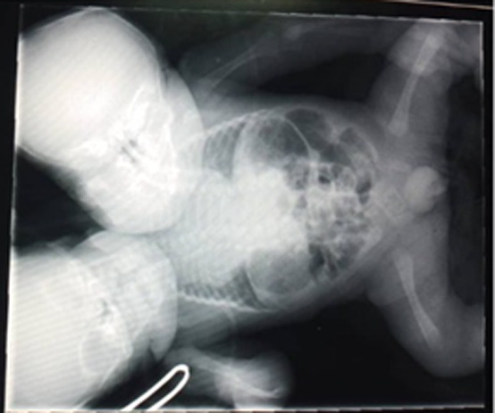
Conjoined twins are a rare occurrence, affecting approximately 1.47 per 100,000 live births as a result of the incomplete splitting of the embryo in monochorionic monoamniotic pregnancies, after 13-14 days post-fertilization. Early prenatal diagnosis through ultrasound between 11 and 14 weeks is crucial for counseling and management decisions, including pregnancy termination or continuation with the support of a multidisciplinary team. We report a case of a 17-year-old primigravida referred in the third trimester with an ultrasound diagnosis of parapagus dicephalus dipus tribrachius conjoined twins. Delivery was by elective cesarean section, and the twins shared a single heart with three ventricles, two atria, a large atrial septal defect, and an incompetent atrioventricular valve. They died of sepsis on day 20 post-delivery. In our setting, anomaly scans during the second trimester are rarely or never performed, highlighting the need for routine early anomaly scans to enable timely intervention and improve outcomes in similar cases.
Keywords: Conjoined twins; case report; congenital anomalies; monoamniotic monochorionic; parapagus dicephalus.
Copyright: Mwaba Kopa et al.
Conflict of interest statement
The authors declare no competing interests.
Figures

References
-
- Farzaneh M, Khoshnam S, Nokhbatolfoghahai M. First scientific record of two cases of partial twinning in the chick embryo, Gallus gallus domesticus. Vet Rec Case Rep. 2017 Feb;4(2):e000353.
-
- Gothwal M, Sharma C, Yadav G, Singh P, Raikar S. Dicephalus parapagus conjoined twin: a rare case with review of literature. Int J Reprod Contracept Obstet Gynecol. 2018;7(8):3410–3412.
-
- Nath S, Munkonge L. Conjoined twins in Zambia. J R Coll Surg Edinb. 1996 Aug;41(4):250–4. - PubMed
Publication types
MeSH terms
LinkOut - more resources
Full Text Sources
Medical
