

The synergy of tuf gene sequencing and maximum likelihood phylogenetic model: a suitable method for identifying the source of coagulase-negative staphylococci infections
- PMID: 40567443
- PMCID: PMC12188015
- DOI: 10.11604/pamj.2025.50.53.44554
The synergy of tuf gene sequencing and maximum likelihood phylogenetic model: a suitable method for identifying the source of coagulase-negative staphylococci infections
Abstract
Introduction: despite ongoing efforts, the health burden of neonatal infection remains unacceptably high due to major challenges, including the hurdle of determining the origin of infection. In this study, we explored the combination of tuf gene sequencing and the Maximum Likelihood Phylogenetic Model (MLPM) as a possible method for investigating the source(s) of transmission of two staphylococcal species, S. epidermidis and S. haemolyticus to neonates and young infants in the Ho Teaching Hospital (HTH) of Ghana, where we previously identified bloodstream infections as a major cause of neonatal morbidity and mortality.
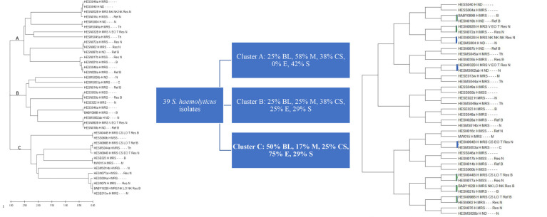
Methods: a total of 106 bacterial isolates were analyzed, comprising 67 S. epidermidis and 39 S. haemolyticus, cultured from blood samples of neonates and young infants, nasal mucosae of mothers, clinical staff, students, and a few objects in the hospital. Isolates were identified using Bruker Daltonik MALDI-TOF, and their nucleic acids were obtained. The tuf genes were sequenced using the Sanger method, and bioinformatics analyses were performed using the MEGA5 (10.1.8 version).
Results: from our data, the combined use of bacterial tuf gene sequencing and MLPM revealed that mothers are the main source of S. haemolyticus-associated neonatal infections, whereas clinical staff is more likely to transmit S. epidermidis to neonates and young infants in the HTH. Whole genome sequencing scatter plots of a few of the isolates were used as a comparator method.
Conclusion: overall, our findings suggest that using bacterial tuf gene sequencing in conjunction with bioinformatic analysis of this gene utilizing the MLPM may serve as a useful epidemiologic method in predicting the source of staphylococcal infections in neonates in the Neonatal Intensive Care Unit (NICU) and possibly other units in the hospital.
Keywords: Tuf gene sequencing; clinical epidemiology; maximum likelihood phylogenetic model; method; neonates; staphylococci(cal) infections.
Copyright: Innocent Afeke et al.
Conflict of interest statement
The authors declare no competing interests.
Figures


References
-
- Sass L, Karlowicz MG. Healthcare-Associated Infections in the Neonate: Principles and Practice of Pediatric Infectious Diseases. 2018:560–566.e3.
MeSH terms
Substances
LinkOut - more resources
Full Text Sources
Medical
