Review
. 2025 Jun 11:13:1547582.
doi: 10.3389/fcell.2025.1547582. eCollection 2025.

KRAS and NRF2 drive metabolic reprogramming in pancreatic cancer cells: the influence of oxidative and nitrosatice stress

Affiliations
Review

KRAS and NRF2 drive metabolic reprogramming in pancreatic cancer cells: the influence of oxidative and nitrosatice stress

Valentina Rapozzi et al. Front Cell Dev Biol. .

Abstract

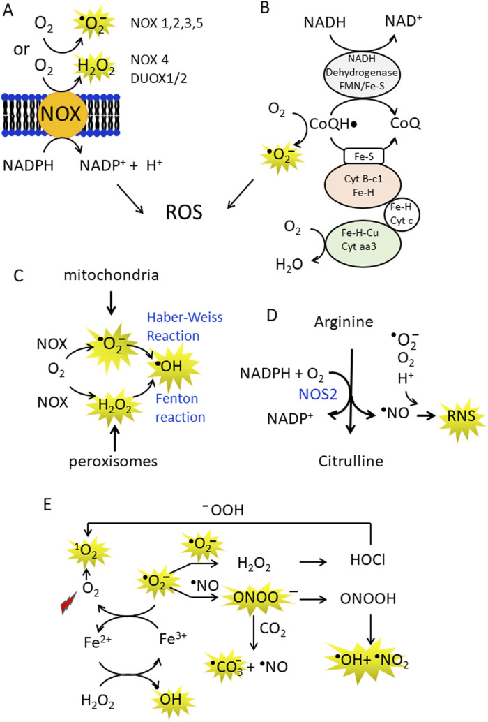
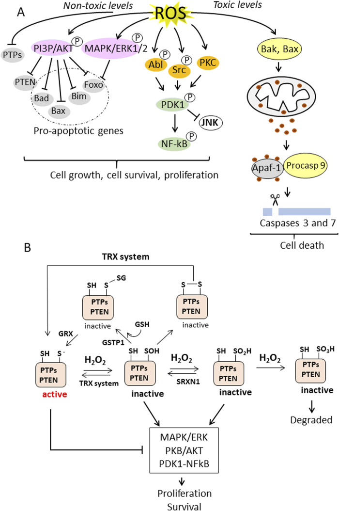
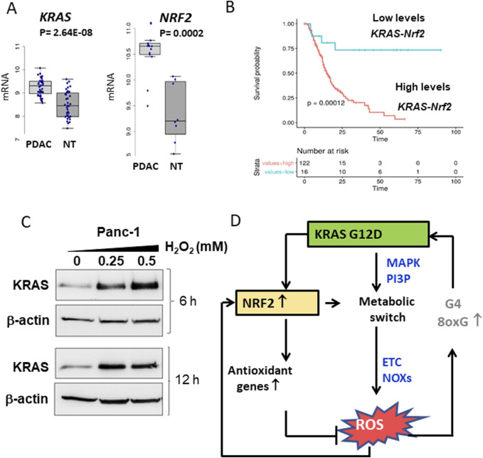
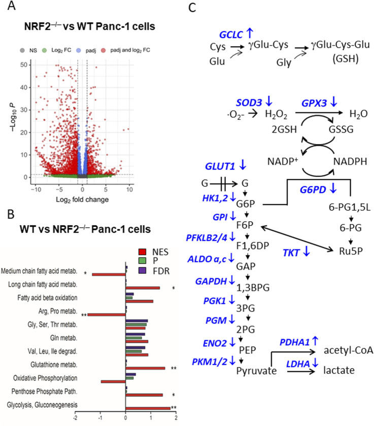
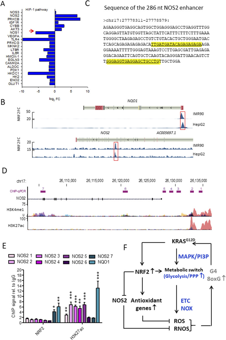
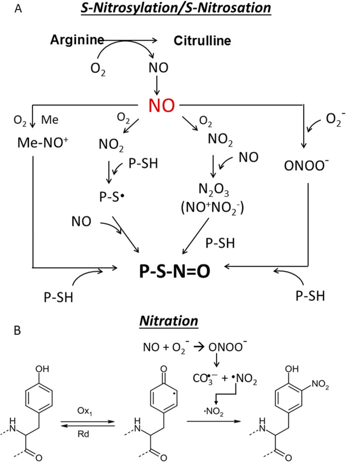
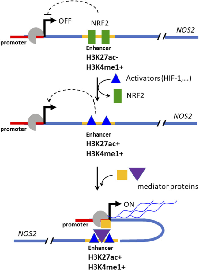
Cancer cells are subject to metabolic reprogramming, which leads to a sustained production of reactive oxygen species (ROS). Increased oxidative stress contributes to genomic instability and promotes malignant transformation. To counteract excessive ROS levels, cells activate nuclear factor erythroid 2-related factor 2 (NRF2), a key regulator of redox homeostasis that coordinates the transcription of a wide range of antioxidant and cytoprotective genes. This review examines the metabolic adaptations controlled by the KRAS-NRF2 axis under oxidative stress conditions. In addition, we highlight a novel function of NRF2 in regulating the expression of NOS2 by binding to a DNA enhancer element, thereby modulating the production of reactive nitrogen species (RNS). Finally, we discuss novel molecular strategies aimed at disrupting adaptive antioxidant responses in cancer cells and provide insights into combinatorial therapeutic approaches targeting redox balance in cancer.

Keywords: KRAS; NOS2; NRF2; PDAC; RNS; ROS; metabolic reprogramming.


Conflict of interest statement

The authors declare that the research was conducted in the absence of any commercial or financial relationships that could be construed as a potential conflict of interest. The author(s) declared that they were an editorial board member of Frontiers, at the time of submission. This had no impact on the peer review process and the final decision.

Figures

FIGURE 1
FIGURE 1
(A,B) Production of superoxide anions (•O2¯) and hydrogen peroxide (H2O2) by NADPH oxidases (NOX enzymes) and CoQ in the mitochondrial electron transport chain (ETC.). In the ETC some electrons are inadvertently transferred from CoQH to O2 in the mitochondria to generate •O2¯; (C) •O2¯ can be non-enzymatically converted to the more reactive hydroxyl radical (•OH) in the Haber-Weiss reaction. In addition, metal ions such as Fe2+, Cu+ can act as single electron donors in the Fenton reaction to give •OH; (D) Arginine is the substrate of NOS2, which converts arginine to citrulline, releasing NO and RNS; (E) A conversion between ROS and RNS occurs in the cell. At high concentrations, NO can combine non-enzymatically with •O2¯ and form peroxynitrite (ONOO¯). Peroxynitrite is a strong oxidising agent that is stable and can diffuse through membranes and interact with proteins (methionine and -SH groups). It can also split into the hydroxyl radical and nitrogen dioxide (NO2). In addition, ONOO¯ can interact with CO2 to form the carbonate anion and NO. The superoxide anion can be converted into H2O2and O2 by spontaneous or enzymatically controlled dismutation. Hydrogen peroxide via Fenton reaction is transformed in •OH or to hypochlorous acid (HClO) by myeloperoxidase. HClO produces singlet oxygen, a strong oxidising agent, in the presence of hydroperoxides.
FIGURE 2
FIGURE 2
(A) ROS-induced cellular signalling in cancer cells. Non-toxic levels of ROS induce the phosphorylation and activation of PI3P/AKT and MAPK/ERK1/2 and the simultaneous inactivation of protein tyrosine phosphatases (PTPs) and lipid phosphatase, resulting in the inhibition of proapoptotic genes and the stimulation of cell growth and survival. ROS also activates the PDK1/NF-kB signalling pathway, leading to survival and proliferation. In contrast, overproduction of ROS leads to toxic oxidative stress, which activates Bak (Bcl-2 homologue antagonist/killer) and Bax (Bcl-2-associated X protein), which are pro-apoptotic members of the Bcl-2 protein family that regulate apoptosis and in particular the intrinsic (mitochrondrial) apoptosis pathway. Bax and Bak form pores in the outer mitochrondrial membrane that allow the release of cytochrome c into the cytoplasm. Cytochrome c induces the formation of the apoptosome, which activates caspase 9, which in turn activates executioner caspases 3 and 7; (B) PTPs and PTEN have a Cys residue in the active site in a thiolate state, which is susceptible to oxidation. The thiolate can be oxidised to sulfenate (-SOH), sulfinate (-SO2H) or sulfonate (-SO3H) depending on the H2O2 concentration. The Cys in the active site can also form disulfides, either with GSH, a reaction catalysed by GSTP1, or with another thiol in the active site, forming a disulphide bridge. These oxidative modifications inactivate the phosphatases and thereby enhance the MAPK/ERK and PKB/AKT pathways. The oxidative inactivation to sulfenate or sulfinate can be reversed by the antioxidant systems TXN1 or SRXN1. This restores phosphatase activity and promotes suppression of the MAPK/ERK and PKB/AKT signalling pathways. Similarly, Cys in the active site that have formed S-glutathionylation or have formed a disulfide bridge can be reversed by the enzyme glutaredoxin (GRX) (catalyses the reversible reduction of glutathione-protein mixed disulfide) or the TRX system. The oxidation of Cys in the active centre to a sulfonate state is irreversible (right) and the altered protein is degraded.
FIGURE 3
FIGURE 3
(A) Differential expression of KRAS and NRF2 between normal and tumor tissues in PDAC patients. Data obtained from GSE15471; (B) Kaplan-Meir plots show that patients with high levels of KRAS and NRF2 expression exhibit a lower survival probability than patients with low KRAS and NRF2 expression; (C) Levels of KRAS and actin in Panc-1 cells treated with increasing amounts of H2O2; (D) Scheme showing the relationships between KRAS, NRF2 and ROS in pancreatic cancer cells. Panels A,B,C adapted with permission (iScience 2023, 26, 108566).
FIGURE 4
FIGURE 4
(A) Volcano plot of DEGs in Panc-1 NRF2−/− cells compared to WT cells; (B) Functional enrichment analysis of WT Panc-1 cells compared to NRF2−/− Panc-1 cells. This analysis shows that in cells where NRF2 was deleted (NRF2−/−), glycolysis, PPP, glutathione metabolism and long-chain fatty acid metabolism are inhibited, while arginine/proline and medium fatty acid metabolism are activated. NES = normal enrichment score; P = p-value; FDR = false discovery rate; (C) Glycolytic, PPP and glutathione metabolism enzymes are downregulated in NRF2−/− cells (indicated with ↓). Panels A and B adapted with permission (iScience 2023, 26, 108566).
FIGURE 5
FIGURE 5
(A) Gene expression of the HIF pathway in NRF2−/− cells. Note that NOS2 is highly expressed in NRF2−/− cells while NOS1 is downregulated. The opposite holds for WT Panc-1 cells; (B) NRF2 ChIP-seq signals expressed as fold-change (FC) respect to Input in correspondence of NQO1 and NOS2 genomic loci. Data were retrieved from Encode and the significantly enriched peaks are highlighted; (C) Sequence of the NOS2 enhancer, the two predicted NRF2 binding sites are highlighted in yellow; (D) H3K4me1 and H3K27ac ChIP-seq signals expressed as fold-change (FC) compared to Input in correspondence of NOS2. The amplified regions investigated in qPCR are indicated (from 1 to 7), H3K4me1 and H3K27ac levels were obtained from Encode (cell lines: GM12878, H1-hESC, HSMM, HUVEC, K562, NHEK, NHLF); (E) ChIP signals relative to IgG obtained with anti-NRF2 and anti-H3K27ac antibodies in WT Panc-1 cells relative to the indicated genomic loci; (F) Interplay between KRAS, NRF2 and NOS2 in the control of oxidative and nitrosative homeostasis in PDAC. Panels A,B,C,D,E adapted with permission (BBA MCR 2024, 1871, 119106).
FIGURE 6
FIGURE 6
(A) S-nitrosylation/S-nitrosation. This cellular modification can occur in different ways. First, NO can form with O2 dinitrogen trioxide N2O3 which isomerises to nitrosonium nitrite (NO+NO2 ) (Zakharov and Zakharov, 2009) whose nitrosonium NO+ reacts with a protein thiol (P-SH) to produce a nitrosothiol (P-S-N=O). In another pathway NO is oxidized to NO2 which reacts with a thiol group to give a thiol radical (-S∙) that with NO provides a nitrosothiol. The third pathway is mediated by metal which generates with NO a nitrosonium (NO+) (Mn+1 + NO + O2→ Mn-NO+), that adds to the thiol group to form a nitrosothiol (Aboalroub and Al Azzam, 2024; Ye et al., 2022; Reactive cysteines of proteins and glutathione (GSH) can undergo S-nitrosation by the peroxynitrite ONOO¯ to form S-nitrosothiol derivatives, P-SNOs and S-nitrosoglutathione (GSNO), respectively. Not all Cys are susceptible to S-nitrosylation. Those that are subject to S-nitrosylation lie within a consensus sequence that includes amino acids that create a hydrophobic environment. S-nitrosylation depends on several factors, including the acid/base and hydrophobic residues in the vicinity of the cysteine and the accessibility of the solvent (Marino and Gladyshev, 2010; Doulias et al., 2010). Notably, a hydrophobic environment attracts hydrophobic gases like NO and O2 and strongly enhances the rate of S-nitrosylation (Möller et al., 2007). S-nitrosation refers to a chemical process that occurs under physiological and pathological conditions in which peroxinitrite ONOO reacts non-enzymatically with the thiol group to form a nitrosothiol (P-S-N=O); (B) Nitration. The one-electron oxidation of tyrosine produces a tyrosine radical Tyr, which is converted into 3-nitrotyrosine by reaction with NO2. As the pK value of the −OH group of Tyr is 10.3, it is 100% protonated at physiological pH, while the pK value drops to 7.3 during nitration and the -OH group is almost 50% deprotonated. This can strongly influence the structure of the nitrated protein.
FIGURE 7
FIGURE 7
Mechanism by which NRF2 suppresses the expression of NOS2 in Panc-1 cells. At elevated concentrations, NRF2 binds to a distal enhancer, inactivating it. The enhancer bound by NRF2 is locked to the activators protein and the expression of NOS2 is inhibited (in this state the DNA at the NOS2 locus is marked by H3K27ac- and H3K5me1+). When the NRF2 level decreases, the enhancer is unlocked, free of NRF2, and is bound by activator proteins that promote transcription (in this state the DNA at the NOS2 locus is marked by H3K27ac+ and H3K5me1+). (“Med” stands for Mediator, a protein complex involved in gene expression in eukaryotic cells; “ac” = activator protein, TSS = transcription start site. Adapted with permission (BBA MCR 2024, 1871, 119106).

Similar articles

References

    1. Abman S. H. (2013). Inhaled nitric oxide for the treatment of pulmonary arterial hypertension. Handb. Exp. Pharmacol. 218, 257–276. 10.1007/978-3-642-38664-0_11 - DOI - PubMed
    1. Aboalroub A. A., Al Azzam K. M. (2024). Protein S-nitrosylation: a chemical modification with ubiquitous biological activities. Protein J. 43, 639–655. 10.1007/s10930-024-10223-y - DOI - PubMed
    1. Anand P., Stamler J. S. (2012). Enzymatic mechanisms regulating protein S-nitrosylation: implications in health and disease. J. Mol. Med. Berl. 90 (3), 233–244. 10.1007/s00109-012-0878-z - DOI - PMC - PubMed
    1. Back E., Coolens K., Van den Bossche J. L., Houbracken I., Espinet E., Rooman I. (2022). On the origin of pancreatic cancer: molecular tumor subtypes in perspective of exocrine cell plasticity. Cell. Mol. Gastroenterol. Hepatol. 13 (4), 1243–1253. 10.1016/j.jcmgh.2021.11.010i - DOI - PMC - PubMed
    1. Bandookwala M., Sengupta P. (2020). 3-Nitrotyrosine: a versatile oxidative stress biomarker for major neurodegenerative diseases. Int. J. Neurosci. 130 (10), 1047–1062. 10.1080/00207454.2020.1713776 - DOI - PubMed

LinkOut - more resources