Case Reports
. 2025 Jun 11:15:1531668.
doi: 10.3389/fonc.2025.1531668. eCollection 2025.

Case Report: A very rare case of diffuse large B-cell lymphoma with cardiac and ovarian involvement

Affiliations
Case Reports

Case Report: A very rare case of diffuse large B-cell lymphoma with cardiac and ovarian involvement

Yadan Du et al. Front Oncol. .

Abstract

Background: Diffuse large B-cell lymphoma (DLBCL) is the most prevalent type of aggressive lymphoma, commonly spreading to sites such as the lymph nodes, spleen, bone marrow, liver, lungs, and central nervous system. However, metastasis to the heart and ovaries is relatively uncommon.

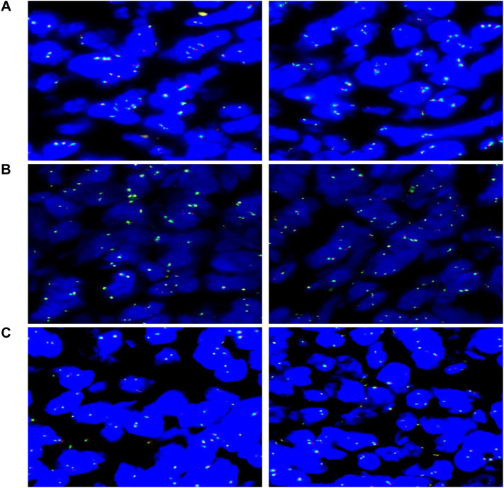
Case description: A 63-year-old woman visited the hospital with abdominal pain and bloating, but showed none of the typical signs of lymphoma. Imaging scans revealed abnormal masses in both the pericardium and ovaries. A biopsy confirmed it was DLBCL, presenting in the rare form of simultaneous spread to the heart lining and ovaries. During the course of illness, she also developed atrial arrhythmia. Doctors adopted a phased treatment approach: four cycles of R-CEOD chemotherapy led to a noticeable reduction in the heart tumor and improvement in her heart rhythm. This was followed by four cycles of R-CHOP, which further shrank the cardiac lesion and cleared the abdominal tumors completely. The treatment was well tolerated, and at a three-month follow-up, there was no sign of recurrence. Her heart function remained stable, with a left ventricular ejection fraction (LVEF) of 60%.

Conclusion: This case highlights the importance of early detection of atypical metastases in DLBCL through a combination of various imaging and pathological tests. Additionally, personalized treatment strategies may contribute to better patient outcomes.

Keywords: case report; diffuse large B-cell lymphoma; heart; non-Hodgkin lymphoma; ovary.


Conflict of interest statement

The authors declare that the research was conducted in the absence of any commercial or financial relationships that could be construed as a potential conflict of interest.

Figures

Figure 1
Figure 1
Serial echocardiographic surveillance of cardiac lesion: (A) Initial transthoracic echocardiography revealed a roughly oval-shaped filling defect within the right atrium, measuring approximately 43 × 34 mm. The lesion appeared to be anchored to the superior aspect of the right atrial wall. It demonstrated well-defined margins and minimal mobility, suggestive of a space-occupying mass with low dynamic activity. (B) Following four cycles of R-CEOD chemotherapy, reevaluation revealed a 14 × 8 mm hypoechoic-to-isoechoic lesion along the posterolateral wall of the right atrium. The lesion remained well-circumscribed, with a near-oval contour and persistently low mobility. (C) After an additional four cycles of R-CHOP, imaging showed the lesion to be stable in size at 19 × 8 mm. Its echogenicity remained mixed hypoechoic, and morphological characteristics (including its oval shape, sharp margins, and low mobility) were consistent with previous findings. The lesion continued to localize to the posterolateral wall of the right atrium. (D) At a three-month post-chemotherapy follow-up, the lesion measured 18 × 7 mm. Its acoustic properties and morphological features (mixed hypoechoic pattern, oval shape, well-defined borders, and limited mobility) remained unchanged. The basal attachment site was concordant with prior assessments.
Figure 2
Figure 2
Radiological evaluation of disease dynamics: (A) Baseline imaging revealed marked enlargement of the right atrial chamber. A lobulated, mixed-density mass measuring approximately 6.5 × 5.6 cm was identified at the junction of the right atrium and pericardium. The lesion exhibited well-demarcated borders and demonstrated pronounced heterogeneous enhancement on contrast-enhanced scans. (B) Initial pelvic imaging identified bilateral adnexal masses characterized by well-defined mixed densities. The largest lesion measured 42.4 × 73.5 × 50.0 mm. Dynamic contrast-enhanced imaging revealed a pattern of persistent enhancement, interspersed with multiple non-enhancing patchy areas throughout the lesion. (C) Post-standard chemotherapy re-evaluation showed significant regression of the primary right atrial lesion, with a maximal axial dimension of 30 × 25 mm. Imaging characteristics were consistent with a localized filling defect. (D) At the final post-chemotherapy pelvic follow-up, complete resolution of bilateral adnexal lesions was observed. No abnormal enhancement or space-occupying lesions were detected. The imaging data comprehensively captured the temporal evolution of lesion morphology and enhancement characteristics throughout the treatment course.
Figure 3
Figure 3
Pathological slide: Diffuse large B-Cell Lymphoma (DLBCL, NOS, Non-GCB subtype).
Figure 4
Figure 4
Molecular pathology diagnostic report: (A) Analysis of 100 tumor cells shows that 7% exhibit abnormal signal patterns, remaining below the 15% threshold, indicating no MYC gene rearrangement (negative). (B) Analysis of 100 tumor cells shows that 4% exhibit abnormal signal patterns, remaining below the 15% threshold, indicating no BCL6 gene rearrangement (negative). (C) Analysis of 100 tumor cells shows that 3% exhibit abnormal signal patterns, remaining below the 15% threshold, indicating no BCL2 gene rearrangement (negative).
Figure 5
Figure 5
Electrocardiogram. (A) Atrial escape rhythm (36 bpm) and junctional escape rhythm were observed. The QT interval was prolonged (525 ms). ST segment depression of 0.05–0.1 mV was noted in leads V2–V4. RV5 amplitude was elevated at 1.28 mV, with a combined RV5 + SV1 value of 1.88 mV. The P wave duration was 82 ms, and the QRS complex measured 94 ms. (B) Sinus rhythm was restored (80 bpm). The QT interval normalized to 410 ms, and the ST segment changes resolved. RV5 amplitude decreased to 0.9 mV, with a combined RV5 + SV1 of 1.57 mV. The P wave measured 74 ms, and the QRS complex was 84 ms.

Similar articles

References

    1. Wang SS. Epidemiology and etiology of diffuse large B-cell lymphoma. Semin Hematol. (2023) 60:255–66. doi: 10.1053/j.seminhematol.2023.11.004 - DOI - PMC - PubMed
    1. Li S, Young KH, Medeiros LJ. Diffuse large B-cell lymphoma. Pathology. (2018) 50:74–87. doi: 10.1016/j.pathol.2017.09.006 - DOI - PubMed
    1. Ollila TA, Olszewski AJ. Extranodal diffuse large B cell lymphoma: molecular features, prognosis, and risk of central nervous system recurrence. Curr Treat options oncology. (2018) 19:38. doi: 10.1007/s11864-018-0555-8 - DOI - PMC - PubMed
    1. Chen DB, Shen DH, Zhang H, Wang Y, Song QJ, Yang SM, et al. Tumors of lymphoid and hematopoietic tissue of spleen: a clinicopathologic analysis of 53 cases. Zhonghua bing li xue za zhi = Chin J pathology. (2017) 46:775–81. doi: 10.3760/cma.j.issn.0529-5807.2017.11.008 - DOI - PubMed
    1. Papanastasiou L, Skarakis N, Vardaka M, Dimitriadi A, Lampropoulou P, Perpinia A, et al. A very rare case of extranodal B-cell non-Hodgkin lymphoma presenting with adrenal and heart involvement. Leukemia Res reports. (2022) 17:100318. doi: 10.1016/j.lrr.2022.100318 - DOI - PMC - PubMed

Publication types

LinkOut - more resources