[Preprint]. 2025 Apr 19:2025.04.17.649426.
doi: 10.1101/2025.04.17.649426.

An optogenetic toolkit for robust activation of FGF, BMP, & Nodal signaling in zebrafish

Affiliations

An optogenetic toolkit for robust activation of FGF, BMP, & Nodal signaling in zebrafish

Leanne E Iannucci et al. bioRxiv. .

Abstract

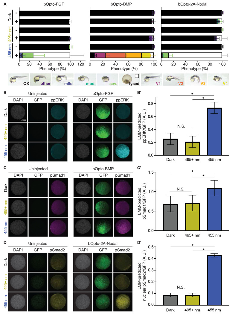
Cell signaling regulates a wide range of biological processes including development, homeostasis, and disease. Accessible technologies to precisely manipulate signaling have important applications in basic and translational research. Here, we introduce an optogenetic toolkit comprised of 1) a zebrafish-optimized FGF signaling activator, 2) a single-transcript Nodal signaling activator, and 3) a previously established BMP signaling activator. We thoroughly characterize this suite of tools in zebrafish embryos and show that they provide tunable, light-dependent spatiotemporal control of signaling in vivo. In response to blue light (~455 nm), receptor kinase domains fused to blue light-dimerizing LOV domains enable robust signaling activation with minimal ectopic activity in the dark or at wavelengths over 495 nm. Optogenetic activation by each tool is pathway-specific and results in increased expression of known target genes. Signaling is activated with rapid on/off kinetics, and activation strength depends on light irradiance. Finally, we demonstrate spatially localized signaling activation with our optimized FGF activator. Together, our results establish this optogenetic toolkit as a potent experimental platform to rapidly, directly, and adjustably activate FGF, BMP, and Nodal signaling in zebrafish embryos.

Keywords: BMP; FGF; Nodal; embryogenesis; molecular optogenetics; signaling; zebrafish.


Conflict of interest statement

Competing interests: No competing interests declared.

Figures

Figure 1:
Figure 1:. Optogenetic signaling activator toolkit.
A,B,C) Schematic of constructs used here to activate FGF (A), BMP (B), and Nodal (C) signaling. Myr = myristoylation motif, GS = glycine/serine linker, LOV = light oxygen voltage-sensing domain, HA = hemagglutinin tag, FLAG = FLAG epitope. The single-transcript bOpto-2A-Nodal construct (C) follows the same design as -FGF and -BMP, except the type I (Acvr1ba) and type II (Acvr2ba) components are connected via a 2A peptide sequence (gray). A’,B’,C’) Optogenetic strategy to activate FGF (A’), BMP (B’), and Nodal (C’) signaling. Blue light-dimerizing LOV domains are fused to myristoylated receptor kinase domains. Blue light exposure should lead to receptor kinase interactions, signaling effector phosphorylation, and activation of target genes.
Figure 2:
Figure 2:. Wavelength-dependent activation of FGF, BMP, & Nodal signaling.
A) Uninjected (−) embryos and embryos injected (+) at the one-cell stage with the indicated mRNA were exposed to dark, 495+ nm light (18.51 W/m2), or 455 nm light (50 W/m2) starting ~2 hours post-fertilization (hpf). Phenotypes were scored at 1 day post-fertilization (dpf) (N = 3; Supp. Fig. 2; Mean +/− SD). B,C,D) Uninjected embryos and embryos injected with the indicated bOpto + GFP mRNA were exposed to dark, 495+ nm light, or 455 nm light starting at early gastrulation (50% epiboly - shield) for 30 min. HCR-IF was used to detect phosphorylated signaling effectors (ppERK, pSmad1, and pSmad2 reflect FGF, BMP, and Nodal signaling, respectively). Scale bar is 200 μm. B’,C’,D’) Quantification of experiments shown in B,C,D. Linear-mixed model-predicted least squared means of GFP-normalized phosphorylated effector signal +/− SEM. D’ shows nuclear signal only. (N = 3, * indicates p < 0.05; Supp. Figs. 3 & 4).
Figure 3:
Figure 3:. bOpto tools activate target gene expression.
Uninjected embryos (−) and embryos injected (+) at the one-cell stage with mRNA encoding bOpto-FGF (A), bOpto-BMP (B), or bOpto-2A-Nodal (C) were exposed to 455 nm light (50 W/m2, bottom rows) or dark (top rows) for 2 hours starting before gastrulation (dome - 30% epiboly). Multiplexed HCR-FISH was used to detect expression of the indicated pathway target genes. (N = 3; Scale bar is 200 μm).
Figure 4:
Figure 4:. Pathway-specific optogenetic activation of FGF, BMP, & Nodal signaling.
Uninjected embryos (−) and embryos injected (+) at the one-cell stage with mRNA encoding bOpto-FGF (A), bOpto-BMP (B), or bOpto-2A-Nodal (C) were exposed to dark (left panel) or 455 nm light (50 W/m2, right panel) for 30 minutes starting at gastrulation (50% epiboly - shield). Triple IF was used to simultaneously detect phosphorylated signaling effectors (ppERK, pSmad1, and pSmad2 reflect FGF, BMP, and Nodal signaling, respectively). (N = 3; Supp. Fig. 5; Scale bar is 200 μm).
Figure 5:
Figure 5:. On/off kinetics of optogenetic signaling activator toolkit.
Embryos were injected at the one-cell stage with mRNA encoding GFP and either bOpto-FGF, bOpto-BMP, or bOpto-2A-Nodal . A,B,C) Starting at early gastrulation (50% epiboly - shield), embryos were either kept in the dark (top row) or exposed to 455 nm light (50 W/m2) light for 30 min (bottom row) and fixed during and after exposure. HCR-IF was used to detect activated signaling effectors (ppERK, pSmad1, and pSmad2 reflect FGF, BMP, and Nodal signaling, respectively). (N = 3; Scale bar is 200 μm). A’,B’,C’) Quantification of experiments shown in A-C. Signal was GFP-normalized and subtracted against dark time-matched controls (N = 3; each N indicated by matched shape; Supp. Fig. 6 & 7; Mean +/− SD; * indicates p < 0.05 when compared to time = 0 min). A”- C”, A’’’-C’’’) A three-parameter logistic regression was fit to the 0-30 min data (A”-C”) or the >= 30 min data (A’’’-C’’’). (N = 3; Mean +/− SD; Supplementary Materials; solid line represents predicted curve fit +/− 95% CI).
Figure 6:
Figure 6:. Irradiance sensitivity of optogenetic toolkit.
A) Table of irradiance and corresponding light dosage values used in B-D’. B,C,D) Embryos were injected at the one-cell stage with mRNA encoding GFP and either bOpto-FGF (B), bOpto-BMP (C), or bOpto-2A-Nodal (D). Starting at early gastrulation (50% epiboly - shield), embryos were exposed to 455 nm light at the indicated irradiance for 5 min (bOpto-FGF, B) or 25 min (bOpto-BMP and -2A-Nodal, C and D). HCR-IF was used to detect phosphorylated signaling effectors (ppERK, pSmad1, and pSmad2 reflect FGF, BMP, and Nodal signaling, respectively; Scale bar is 200 μm). B’,C’,D’) Quantification of experiments in B-D. Signal was GFP-normalized and subtracted against dark controls. A three-parameter logistic regression was fit to the data. (N = 2-3; each N indicated by matched shape; Mean +/− SD; Supp. Fig. 8 & 9; solid line represents predicted curve fit +/− 95% CI; D’ shows nuclear signal only). Tables indicate goodness of fit (R2), the predicted irradiance (with 95% CI) at which 20 (I20), 50 (I50), and 90% (I90) of the curve’s upper asymptote is reached (Supplementary Materials).
Figure 7:
Figure 7:. Spatially localized FGF signaling activation.
Embryos were injected at the one-cell stage with mRNA encoding the green-to-red photoconvertible fluorescent protein nls-Kaede and bOpto-FGF. At early gastrulation (shield) embryos were either kept in the dark (A) or illuminated locally with 405 and 445 nm confocal lasers (B,B’). HCR-IF was used to detect ppERK. Dotted white line in B’ outlines photoconverted Kaede in embryo shown in B. (Supp. Fig. 10; Scale bar is 200 μm).

References

    1. Ando R., Hama H., Yamamoto-Hino M., Mizuno H. and Miyawaki A. (2002). An optical marker based on the UV-induced green-to-red photoconversion of a fluorescent protein. Proc Natl Acad Sci U S A 99, 12651–12656. - PMC - PubMed
    1. Belacortu Y. and Paricio N. (2011). Drosophila as a model of wound healing and tissue regeneration in vertebrates. Dev Dyn 240, 2379–2404. - PubMed
    1. Benedetti L., Barentine A. E. S., Messa M., Wheeler H., Bewersdorf J. and De Camilli P. (2018). Light-activated protein interaction with high spatial subcellular confinement. Proc Natl Acad Sci U S A 115, E2238–E2245. - PMC - PubMed
    1. Benman W., Berlew E. E., Deng H., Parker C., Kuznetsov I. A., Lim B., Siekmann A. F., Chow B. Y. and Bugaj L. J. (2022). Temperature-responsive optogenetic probes of cell signaling. Nat Chem Biol 18, 152–160. - PMC - PubMed
    1. Bennett J. T., Joubin K., Cheng S., Aanstad P., Herwig R., Clark M., Lehrach H. and Schier A. F. (2007). Nodal signaling activates differentiation genes during zebrafish gastrulation. Dev Biol 304, 525–540. - PMC - PubMed

Publication types

LinkOut - more resources