[Preprint]. 2025 Feb 27:2025.02.21.639368.
doi: 10.1101/2025.02.21.639368.

Functional, Immunogenetic, and Structural Convergence in Influenza Immunity between Humans and Macaques

Affiliations

Functional, Immunogenetic, and Structural Convergence in Influenza Immunity between Humans and Macaques

Maya Sangesland et al. bioRxiv. .

Update in

  • Functional, immunogenetic, and structural convergence in influenza immunity between humans and macaques.
    Sangesland M, Li N, Tsybovsky Y, Rodgers MD, Han J, Rodriguez AJ, Ferguson JA, Henry AR, Smith SC, Roberts-Torres J, Gillespie RA, Liu C, Merriam JS, Stephens T, Williams C, Walker E, Corcoran M, Ravichandran M, Creanga A, Andrews SF, Pierson TC, Karlsson Hedestam GB, Schramm CA, Reed DS, Douek DC, Zhou T, Ward AB, Kanekiyo M. Sangesland M, et al. Sci Transl Med. 2025 Sep 10;17(815):eady3570. doi: 10.1126/scitranslmed.ady3570. Epub 2025 Sep 10. Sci Transl Med. 2025. PMID: 40929243

Abstract

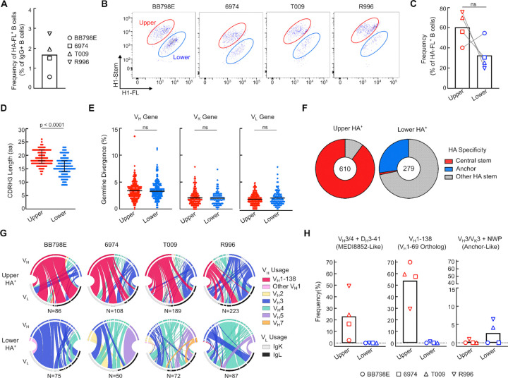
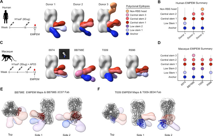
Human B cell immunity to the influenza hemagglutinin (HA) stem region, a universal influenza vaccine target, is often stereotyped and immunogenetically restricted, posing challenges for study outside humans. Here, we show that macaques vaccinated with a HA stem immunogen elicit human-like public B cell lineages targeting two major conserved sites of vulnerability, the central stem and anchor epitopes. Central stem antibodies were predominantly derived from VH1-138, the macaque homolog of human VH1-69, a VH-gene preferentially used in human central stem broadly neutralizing antibodies (bnAbs). Similarly, macaques produced anchor bnAbs with the human-like NWP motif. Both bnAb lineages were functionally and structurally analogous to their human counterparts, with recognition mediated largely by germline-encoded motifs. Thus the macaque immunoglobulin repertoire supports human-like public bnAb responses to influenza HA. Moreover, this underscores the utility of homologous germline-encoded immunity, suggesting that immune repertoires of macaques and humans may have been similarly shaped during evolution.

Keywords: B cell; Nonhuman primates; VH1–69; broadly neutralizing antibody; germline-encoded motif; hemagglutinin; influenza; public lineage; stem nanoparticle.


Conflict of interest statement

DECLARATION OF INTERESTS M.K. is named as an inventor on patents describing the HA stem immunogen used in this study under US10363301, US11147867, US11679151 which were filed by the Department of Health and Human Services.

Figures

Figure 1.
Figure 1.. Serum epitope specificity and neutralizing activity in macaques following H1ssF immunization
(A) Sequential immunization scheme with H1ssF. Influenza naive cynomolgus macaques (n = 7) were vaccinated thrice with H1ssF and AF03 adjuvant. (B) Serum IgG endpoint titer to recombinant H1 (NC99) HA trimeric protein at wks 0, 2, 4, 6, 8, 10 and 12. Arrows denote vaccination timepoints. Horizontal line denotes limit of detection. N = 7 animals, mean and SD are shown. (C) Serum neutralizing antibody response against H1N1 A/New Caledonia/20/1999 (NC99), H1N1 A/Michigan/45/2015 (MI15), H5N1 A/Vietnam/1203/2004 (VN04), and H2N2 A/Singapore/1/1957 (SG57) reporter viruses. Purple line denotes mean. Statistics performed by one-way ANOVA with Geisser-Greenhouse correction and Tukeys post hoc test. (D) Representation of H1 NC99 HA trimer probes: WT HA containing central stem and anchor epitopes (H1-HA), addition of a N45HA2 glycan (red, H1-Δcentral), and addition of a N27HA2 glycan (blue, H1-Δanchor). Central stem (red, PDB: 3GBM) and anchor (blue, PDB: 6HJQ) epitopes are mapped on HA (PDB: 3LZG) (Top). Binding (AUC) of representative head (CH65), central stem (CR9114 and 315–02-1H01), and anchor (FISW84) mAbs against H1-HA, H1-Δcentral, H1-Δanchor probes (Bottom). Samples run in duplicate. (E-F) Serum IgG response at week 12 to H1-HA (black line), H1-Δcentral (red line), and H1-Δanchor (blue line). N=7 animals, mean and SD are shown (E). Corresponding serum binding (AUC) at week 12. Statistical significance determined by one-way ANOVA with Geisser-Greenhouse correction and Tukeys post hoc test (F). (G) Microneutralization (IC50) titer of immune sera at week 12 against H1N1 NC99 and H1N1 MI15 pre-absorbed with the following competitors: none (grey), RSV-F (tan), H1-HA (black), H1-Δcentral (red), H1-Δanchor (blue). Statistical significance determined by one-way ANOVA with Geisser-Greenhouse correction and Tukeys post hoc test (n = 7 animals, line denotes mean).
Figure 2.
Figure 2.. Antigenic and immunogenetic characteristics of HA specific B cells
(A) Frequency of antigen specific B cells reactive to H1-FL at week 12, relative to IgG+ B cells. H1-FL+ B cells are defined as CD3/CD14/CD16/CD56/CD20+/IgG+/IgM/H1-FL+ (see Figure S1D for gating). Symbols denote individual animals. (B-C) Flow plots of HA+ (H1-stem+/H1-FL+) upper cluster (red) and lower cluster (blue) B cells across 4 macaques (B). Corresponding frequency of each upper and lower cluster as a percent of H1-FL reactive B cells (see also Figure S1D for gating). N = 4 animals are shown, symbols denote individual animals. Statistical significance determined by Wilcoxon matched-pairs signed rank test (C). (D) CDR H3 length distribution for upper and lower HA+ clusters. Shown are median and interquartile range. Significance determined by two-tailed Mann-Whitney test. (E) Percent germline divergence across VH, VK and VL chains in each corresponding upper and lower cluster. Significance determined by two-tailed Mann-Whitney test. (F) Antigenicity of upper and lower HA+ sorted single B cells from RATP-Ig supernatants. Antigenicity was determined by binding to H1-HA, H1-Δcentral, and H1-Δanchor probes (see also Figure 1D for probes). Antibody supernatants that did not bind H1-Δcentral or H1-Δanchor probes were denoted as “Other HA stem”. (G) Chord diagrams showing VH (top half) and VL (bottom half) gene pairs in HA+ B cells from the upper and lower clusters. Total number of sequence pairs are listed. (H) Frequency of the relevant lineages; MEDI8852-Like: VH3/4+DH3–41, VH1–69 homolog: VH1–138, and human anchor-like: VH3/VK3+NWP, across 4 macaques (symbols denote individual animals).
Figure 3.
Figure 3.. Macaque central stem and anchor antibodies share functional characteristics to their human counterparts
(A) Microneutralization (IC50) activity for macaque H1ssF elicited monoclonal antibodies against H1N1 A/New Caledonia/20/1999 (NC99), H1N1 A/Michigan/45/2015 (MI15), H5N1 A/Vietnam/1203/2004 (VN04), H2N2 A/Singapore/1/1957 (SG57), and H3N2 A/Wisconsin/67/2005 (WI05) reporter viruses. Central stem (red) and anchor (blue) mAbs are denoted. Bar corresponds to mean. Statistical significance determined by two-tailed Mann-Whitney test. (B) IC50 values of prototypic influenza stem bnAbs. Central stem: 315–02-1H01 (human VH1–69 bnAb) and MEDI8852; Anchor: FISW84; Group 2 stem: CR8020. (C) Characteristics of 3 central stem and 3 anchor macaque bnAbs. Lineage, epitope specificity, and microneutralization titer are shown (see also Figure S2A). (D) Pre-exposure prophylaxis experiment with central stem (BB798E-3C07, BB798E-3A03, and 6974–2G10) and anchor (T009–1H06, T009–3B01, T009–3E04) antibodies. BALB/c mice were treated with 10 mg kg−1 of mAbs via the intraperitoneal route 24 h prior to intranasal virus challenge with H1N1 A/California/04/2009 and H5N1 A/Vietnam/1203/2004 viruses. Percent survival and percent body weight change are shown. Error bars indicate SD. Multiple comparisons of Kaplan-Meier curves were performed by the log-rank test with Bonferroni correction.
Figure 4.
Figure 4.. IGHV1–138 central stem bnAb lineages and structural analysis
(A) Heavy chain genetic characteristics of human IGHV1–69*01 and macaque IGHV1–138 bnAbs. Central stem bnAbs are defined as those that neutralize H1N1 and H5N1 reporter viruses. The corresponding germline alleles and sequences for CDR H1 and CDR H2 are shown as reference. Critical CDR H2 residues at position 53/54 and CDR H3 tyrosine residues are highlighted in red. Kabat numbering is indicated for CDR H1, CDR H2, and CDR H3. (B) Percent germline divergence (nucleotide level) of previously identified human VH1–69 bnAbs compared to macaque VH1–138 bnAbs. P values were calculated using a two-tailed Mann-Whitney test. (C) Cryo-EM map of BB798E-3C07 in complex with H1 NC99 HA trimer. Heavy and light chains of the Fab are colored in red (HC) and light pink (LC). (D) Contact interface between HA (surface representation) and BB798E-3C07. CDR regions of Fab and residues in contact with HA are labeled. The contact area on HA is colored yellow. (E) Detailed illustration of Fab BB798E-3C07 CDR H2 interaction with HA. Only molecular regions participating are shown for clarity. Dashed lines depict hydrogen bonds. (F) Superposition of Fab BB798E-3C07 with the crystal structure of Fab CR6261 (PDB: 3GBM) (Left). Comparison of the contact interface between BB798E-3C07 and CR6261 (Middle). Superposition of residues of BB7998E-3C07 Fab and CR6261 Fab forming key contacts with HA (Right). BB798E-3C07 is shown in red and CR6261 is shown in orchid. (G) Contributions of each CDR to total BSA for BB798E-3C07 and CR6261. Only CDRs participating in the interaction are shown.
Figure 5
Figure 5. Human IGHV1–69 homologs are found across the primate order
(A) Phylogenetic relationship of primate species based on the NCBI taxonomy browser (Left). Percent nucleotide sequence homology of primate IGHV1–69*01 homologs compared to the human IGHV1–69*01 VH-gene (Middle). Corresponding CDR H2 amino acid residues within each primate IGHV1–69 homolog colored by hydrophobicity (orange: high, blue: low) based on the scale of Kyte and Doolittle (Right). (B) BLI sensogram of gHgL BB798E-3C07 ‘CDR H2-swap’ mAbs binding to H1 NC99 HA. Swap mAbs are derived from gHgL BB798E-3C07 where the original macaque CDR H2 domain of BB798E-3C07 is replaced by the CDR H2 sequence from a corresponding VH1–69 primate homolog. All binding curves are compared to macaque gHgL BB798E-3C07. (C) Table comparing the gHgL BB798E-3C07 swap mAbs and their corresponding CDR H2 sequences (see also Figure 5A for CDRH2 sequences). Red denotes residues changed from the original macaque CDR H2 sequence. +/− denotes strength of binding to H1 NC99 HA.
Figure 6.
Figure 6.. Structural characteristics of the anchor bnAb T009–3E04 in complex with HA
(A) Heavy and light chain genetic characteristics of human and macaque anchor antibodies. Critical residues are highlighted in red. Kabat numbering is indicated. (B) Cryo-EM map of Fab T009–3E04 in complex with H1 NC99 HA trimer. Heavy and light chains of Fab are colored in blue (HC) and light blue (LC). (C) Contact interface between HA (surface representation) and T009–3E04. CDR regions of Fab and residues in contact with HA are labeled. The contact area on HA is colored yellow. (D) Detailed illustration of HA and T009–3E04 with participating interactions shown. Dashed lines depict hydrogen bonds and salt bridges. Colors are consistent with panels B and C. (E) Superposition of T009–3E04 with prototypic human anchor antibodies FISW84 (PDB: 6HJQ), 222–1C06 (PDB: 7T3D) and 206–1B06 (PDB: 8D21). Comparison of the contact interface between T009–3E04 (colored in blue) and FISW84, 222–1C06, and 206–1B06 (outlined in green) is shown. (F) CDR Contribution to total BSA for T009–3E04, FISW84, 222–1C06, and 204–1B06. Only participating CDRs are shown.
Figure 7.
Figure 7.. Comparison of polyclonal serum antibody response between humans and macaques vaccinated with H1ssF
(A-D) Electron microscopy polyclonal epitope mapping of serum antibody specificities elicited by H1ssF immunization in humans at week 20 (A-B) and macaques at week 12 (C-D). Central stem epitopes include ‘CR9114-like’ serum mAbs and low stem epitopes include ‘MEDI8852-like’ serum mAbs. (E-F) Overlays of BB798E-3C07 (central stem bnAb) and T009–3E04 (anchor epitope bnAb) cryoEM bnAb models on polyclonal epitope maps of macaque BB798E (E) and T009 (F), respectively.

References

    1. Schroeder H.W. Jr, and Cavacini L. (2010). Structure and function of immunoglobulins. J. Allergy Clin. Immunol. 125, S41–S52. 10.1016/j.jaci.2009.09.046. - DOI - PMC - PubMed
    1. Xu J.L., and Davis M.M. (2000). Diversity in the CDR3 region of V(H) is sufficient for most antibody specificities. Immunity 13, 37–45. 10.1016/s1074-7613(00)00006-6. - DOI - PubMed
    1. Henry Dunand C.J., and Wilson P.C. (2015). Restricted, canonical, stereotyped and convergent immunoglobulin responses. Philos. Trans. R. Soc. Lond. B Biol. Sci. 370. 10.1098/rstb.2014.0238. - DOI - PMC - PubMed
    1. Kwong P.D., and Mascola J.R. (2018). HIV-1 Vaccines Based on Antibody Identification, B Cell Ontogeny, and Epitope Structure. Immunity 48, 855–871. 10.1016/j.immuni.2018.04.029. - DOI - PubMed
    1. Sangesland M., and Lingwood D. (2021). Antibody Focusing to Conserved Sites of Vulnerability: The Immunological Pathways for “Universal” Influenza Vaccines. Vaccines (Basel) 9. 10.3390/vaccines9020125. - DOI - PMC - PubMed

Publication types

LinkOut - more resources