. 2025 May 22;5(1):ycaf074.
doi: 10.1093/ismeco/ycaf074. eCollection 2025 Jan.

Systematic evaluation of protein extraction for metaproteomic analysis of marine sediment with high clay content

Affiliations

Systematic evaluation of protein extraction for metaproteomic analysis of marine sediment with high clay content

Anne Ostrzinski et al. ISME Commun. .

Abstract

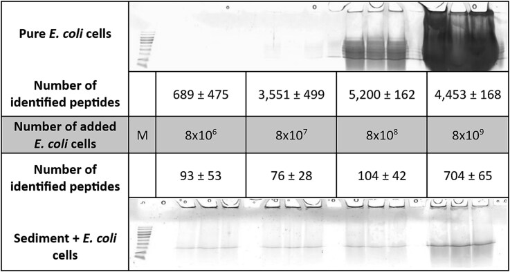
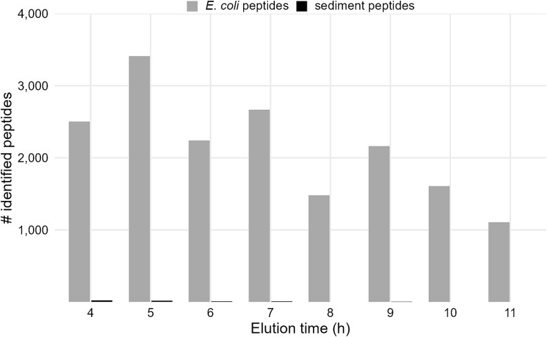
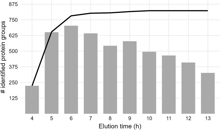
Marine sediments harbor extremely diverse microbial communities that contribute to global biodiversity and play an essential role in the functioning of ecosystems. However, the metaproteome of marine sediments is still poorly understood. The extraction of proteins from environmental samples is still a challenge, especially from marine sediments, due to the complexity of the matrix. Therefore, methods for protein extraction from marine sediments need to be improved. To develop an effective workflow for protein extraction for clayey sediments, we compared, combined and enhanced different protein extraction methods. The workflow presented here includes blocking of protein binding sites on sediment particles with high concentrations of amino acids, effective cell lysis by ultrasonic capture, electro-elution, and simultaneous fractionation of proteins. To test the protocol's efficacy, we added Escherichia coli cells to sediment samples before protein extraction. By using our refined workflow, we were able to identify a comparable number of E. coli proteins from the supplemented sediment to those from pure E. coli cultures. This new protocol will enable future studies to identify active players in clay-rich marine sediments and accurately determine functional biodiversity based on their respective protein complements.

Keywords: Metaproteomics; marine sediment; protein extraction.


Conflict of interest statement

None declared.

Figures

Figure 1
Figure 1
Overview of tested protein extraction steps and optimized extraction workflow. While testing the extraction workflows, all extractions were carried out in triplicates using 5 g of sediment (wet weight). Different numbers of E. coli cells and different concentrations of amino acids were added to the sediment prior to the addition of extraction buffer and heating of the samples. After cell disruption by either ultra-sonication or bead-beating, proteins were separated from the sediment particles by centrifugation or in the BioRad Prep Cell. After TCA precipitation, protein extracts were processed by SDS-PAGE or FASP. The optimized extraction workflow was carried out on 10 g of sediment supplemented with amino acids. After cell disruption via ultra-sonication in urea/thiourea, proteins were extracted from the sediment using the prep cell. Finally, all samples were desalted using C18 ZipTips and measured by LC–MS/MS.
Figure 2
Figure 2
SDS-gels and number of identified peptides obtained from different numbers of E. coli cells (top) or E. coli cells spiked in 5 g of sediment (bottom). Experiments were performed in triplicates, means and standard deviations of identification numbers within replicates were calculated. Samples were mixed with urea/thiourea and cells were disrupted by heating and ultra-sonication. Sediment particles were removed by centrifugation and proteins were enriched by TCA precipitation before SDS-PAGE. Peptide identification was performed as described. M = marker.
Figure 3
Figure 3
Peptides identified in sediment samples treated with different concentrations of an amino acid solution. The experiments were carried out in triplicates, means and standard deviations of identification numbers within replicates were calculated. About 8 × 108  E. coli cells were added to the sediment samples. After the addition of positive polar amino acids solution (25% or 50% AA) or water (no AA), samples were mixed with urea/thiourea, and cells were disrupted by heating and ultra-sonication as described.
Figure 4
Figure 4
Peptides identified according to different strategies for cell disruption and protein processing. The experiments were carried out in triplicates, means and standard deviations of identification numbers within replicates were calculated. Sediment samples were spiked with approximately 8 × 108  E. coli cells. After addition of positive polar amino acids solution (50%), samples were mixed with urea/thiourea. Cells were disrupted with bead-beating or ultra-sonication.
Figure 5
Figure 5
Peptides identified in eluted fractions of the electro-elution approach. The sediment sample was spiked with approximately 8 × 108  E. coli cells. After the addition of positive polar amino acids solution, the sample was mixed with urea/thiourea, and cells were disrupted by heating and ultra-sonication as described. Proteins were electro-eluted from the resulting sediment slurry using the BioRad Prep Cell. After ~4 h, the ion front and the first proteins were eluted.
Figure 6
Figure 6
Number of identified protein groups in eluted fractions. After the addition of positive polar amino acids solution to the sediment, the sample was mixed with urea/thiourea, and cells were disrupted by heating and ultra-sonication. Proteins were electro-eluted from the resulting sediment slurry in the BioRad Prep Cell. Grey bars indicate the number of proteins groups identified in the respective fraction. The black line shows accumulated numbers of proteins groups.
Figure 7
Figure 7
Proportion of assigned phyla and respective functions of identified protein groups. Phyla were determined based on the metagenomic information. Protein functions were determined by EggNOG based on COG categories (Table S2) [46].

References

    1. Hoshino T, Doi H, Uramoto G-I et al. Global diversity of microbial communities in marine sediment. Proc Natl Acad Sci USA 2020;117:27587–97. 10.1073/pnas.1919139117 - DOI - PMC - PubMed
    1. Pradet-Balade B, Boulmé F, Beug H et al. Translation control: bridging the gap between genomics and proteomics? Trends Biochem Sci 2001;26:225–9. 10.1016/S0968-0004(00)01776-X - DOI - PubMed
    1. Wilmes P, Bond PL. Metaproteomics: studying functional gene expression in microbial ecosystems. Trends Microbiol 2006;14:92–7. 10.1016/j.tim.2005.12.006 - DOI - PubMed
    1. Wang D-Z, Xie Z-X, Zhang S-F. Marine metaproteomics: current status and future directions. J Proteome 2014;97:27–35. - PubMed
    1. Saito MA, Bertrand EM, Duffy ME et al. Progress and challenges in ocean metaproteomics and proposed best practices for data sharing. J Proteome Res 2019;18:1461–76. 10.1021/acs.jproteome.8b00761 - DOI - PMC - PubMed

LinkOut - more resources