

Comprehensive examination of resting state fMRI connectomics yields new insights into brain function deficits in Gulf War illness after accounting for heterogeneity in brain impairment across the ill veteran population
- PMID: 40568571
- PMCID: PMC12172930
- DOI: 10.1016/j.ynirp.2024.100209
Comprehensive examination of resting state fMRI connectomics yields new insights into brain function deficits in Gulf War illness after accounting for heterogeneity in brain impairment across the ill veteran population
Abstract
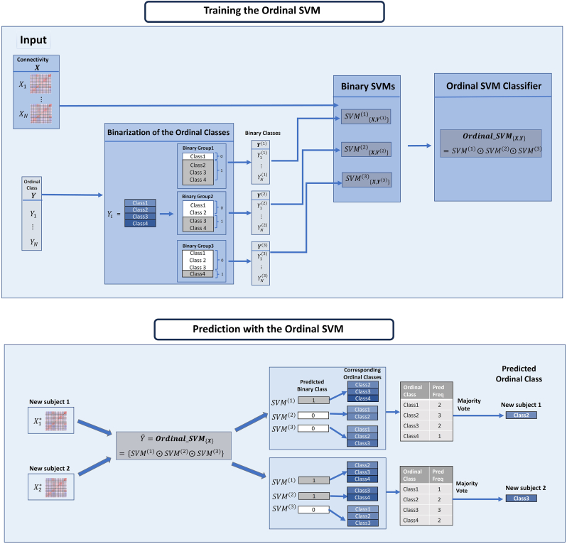
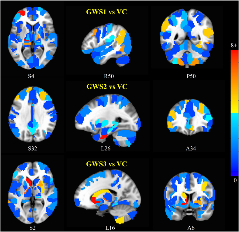
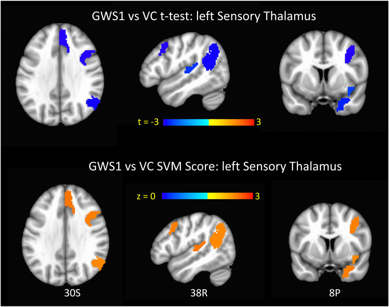
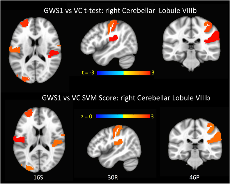
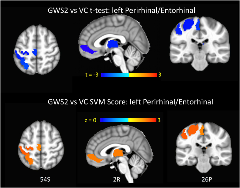
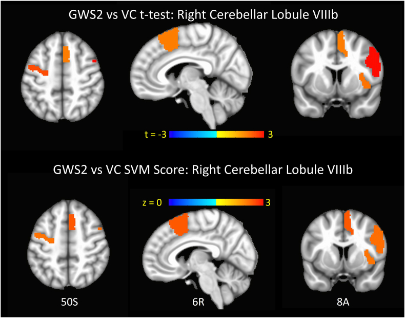
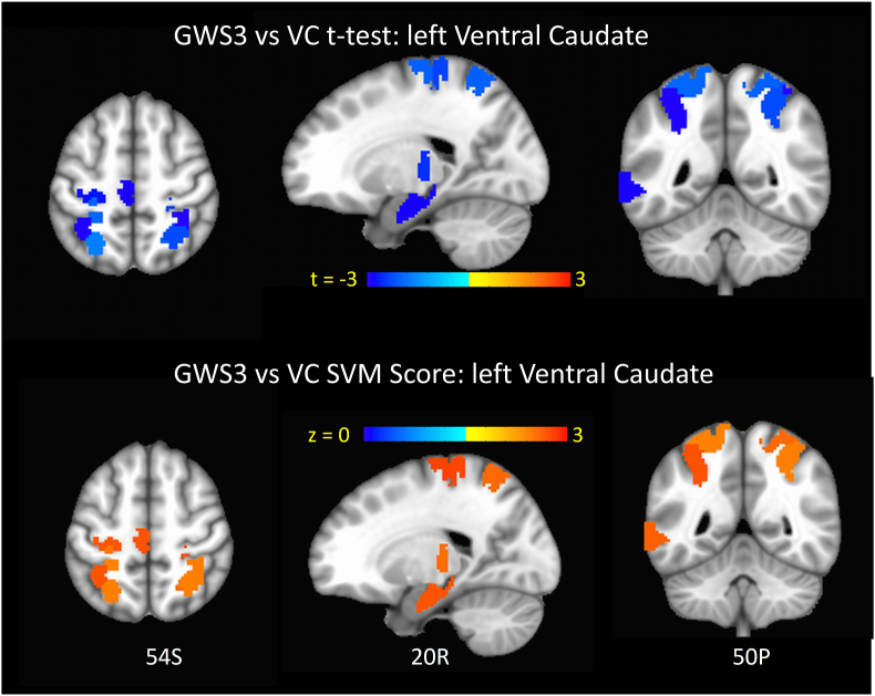
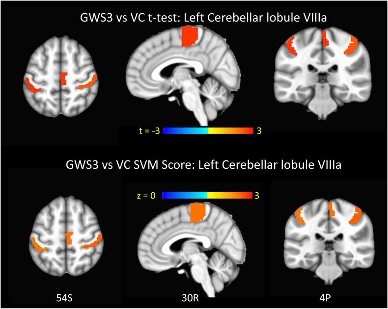
An estimated 200,000 veterans (up to 32% of those deployed) of the 1991 Gulf War (GW) suffer from GW illness (GWI), an incompletely understood chronic medical condition, characterized by multiple symptoms indicative of brain function deficits in various domains. Epidemiologic and animal studies have associated GWI with exposure to neurotoxic chemicals such as nerve agents, organophosphate pesticides and pyridostigmine bromide. One factor that hampers mechanistic investigations into GWI is that there is considerable heterogeneity in brain impairments across the ill GW veteran population. This could reflect the underlying heterogeneity in both exposure to neurotoxic substances, as well as genetic predisposition or resistance to neurotoxicity. Only one of the validated case definitions, the Haley GWI criteria addresses this heterogeneity. It does so by breaking down GWI into three main syndrome variants (GWS1, GWS2, and GWS3) based on factor analysis of symptoms presented by GWI veterans. Resting state fMRI (rsfMRI) is a uniquely useful brain imaging technique in that in a 10-min fMRI scan it can probe numerous brain function domains simultaneously. In this study, we employed a connectomics approach and machine learning on rsfMRI data from a cohort of GW veterans to extract neuroimaging biomarkers specific to each of the three Haley GWI syndromes. Our results revealed a number of new insights into brain function impairment specific to each syndrome group. The findings indicate that these deficits may by and large be driven by brain mechanisms. We also found that pooling the data of all three syndromes in GWI group, as is done by commonly employed case definitions of GWI resulted in failure to detect the fMRI signatures of a lot of these brain impairments.
Keywords: Biomarkers; Brain function networks; Connectomics; Functional connectivity; Gulf war illness; Heterogeneity; Neurotoxic; Ordinal support vector machine; Resting state fMRI.
© 2024 The Authors.
Conflict of interest statement
The authors declare that they have no known competing financial interests or personal relationships that could have appeared to influence the work reported in this paper.
Figures
Similar articles
-
Genetic association between the APOE ε4 allele, toxicant exposures and Gulf war illness diagnosis.Environ Health. 2023 Jul 6;22(1):51. doi: 10.1186/s12940-023-01002-w. Environ Health. 2023. PMID: 37415220 Free PMC article.
-
Anthrax and Gulf War Illness (GWI): Evidence for the Presence of Harmful Anthrax Antigen PA63 In the Serum of Veterans with GWI.J Neurol Neuromedicine. 2019 Nov 25;4(6):1-9. doi: 10.29245/2572.942x/2019/6.1255. J Neurol Neuromedicine. 2019. PMID: 40747435 Free PMC article.
-
In silico Analysis of the Binding Affinities of Antigenic Epitopes of Vaccines Administered to Gulf War Veterans to Specific HLA Class II Alleles Protective for Gulf War Illness.J Neurol Neuromedicine. 2019 Oct 19;4(5):23-30. doi: 10.29245/2572.942x/2019/5.1254. J Neurol Neuromedicine. 2019. PMID: 40746427 Free PMC article.
-
Signs and symptoms to determine if a patient presenting in primary care or hospital outpatient settings has COVID-19.Cochrane Database Syst Rev. 2022 May 20;5(5):CD013665. doi: 10.1002/14651858.CD013665.pub3. Cochrane Database Syst Rev. 2022. PMID: 35593186 Free PMC article.
-
Immunogenicity and seroefficacy of pneumococcal conjugate vaccines: a systematic review and network meta-analysis.Health Technol Assess. 2024 Jul;28(34):1-109. doi: 10.3310/YWHA3079. Health Technol Assess. 2024. PMID: 39046101 Free PMC article.
References
-
- Abdullah L., Evans J.E., Bishop A., Reed J.M., Crynen G., Phillips J., Pelot R., Mullan M.A., Ferro A., Mullan C.M., Mullan M.J., Ait-Ghezala G., Crawford F.C. Lipidomic profiling of phosphocholine-containing brain lipids in mice with sensorimotor deficits and anxiety-like features after exposure to Gulf War agents. NeuroMolecular Med. 2012;14:349–361. - PubMed
-
- Abdullah L., Evans J.E., Montague H., Reed J.M., Moser A., Crynen G., Gonzalez A., Zakirova Z., Ross I., Mullan C., Mullan M., Ait-Ghezala G., Crawford F. Chronic elevation of phosphocholine containing lipids in mice exposed to Gulf War agents pyridostigmine bromide and permethrin. Neurotoxicol. Teratol. 2013;40:74–84. - PubMed
-
- Balazova Z., Novakova M., Minsterova A., Rektorova I. Structural and functional magnetic resonance imaging of dementia with lewy bodies. Int. Rev. Neurobiol. 2019;144:95–141. - PubMed
-
- Batton A.D., Blaha C.D., Bieber A., Lee K.H., Boschen S.L. Stimulation of the subparafascicular thalamic nucleus modulates dopamine release in the inferior colliculus of rats. Synapse. 2018 - PubMed
LinkOut - more resources
Full Text Sources