. 2025 Jun 11:16:1574246.
doi: 10.3389/fimmu.2025.1574246. eCollection 2025.

IL-17A drives a fibroblast-neutrophil-NET axis to exacerbate immunopathology in the lung with diffuse alveolar damage

Affiliations

IL-17A drives a fibroblast-neutrophil-NET axis to exacerbate immunopathology in the lung with diffuse alveolar damage

Duo Su et al. Front Immunol. .

Abstract

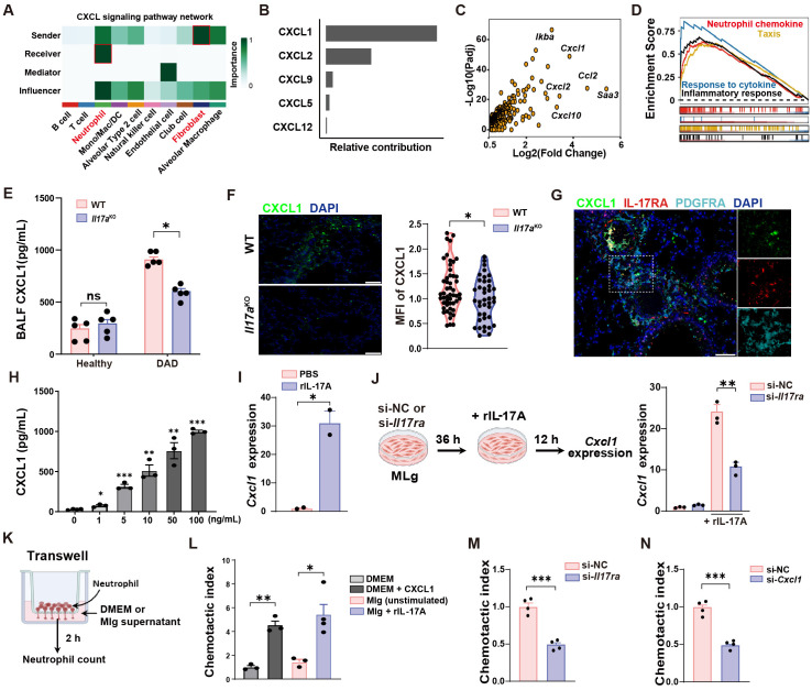
Diffuse alveolar damage (DAD), a lethal manifestation of acute lung injury, remains a critical public health concern due to the absence of targeted therapies. However, the underlying cellular and molecular mechanisms responsible for immunopathology during DAD progression are largely undefined. Here, by integrating single cell RNA sequencing, functional assays, and genetic/pharmacological interventions in a mouse model of ricin-induced DAD, we revealed a significant accumulation of neutrophil with an activated phenotype that plays a critical role in immunopathology. We observed the formation of neutrophil extracellular traps (NETs) during DAD, which further intensified inflammation and tissue injury. IL-17A signaling activity was upregulated in DAD-affected lungs, while IL-17A deficiency or functional blockade significantly attenuated neutrophil recruitment, NET generation, and tissue damage. Mechanically, IL-17A stimulates lung resident fibroblasts to produce the neutrophil chemoattractant CXCL1. Notably, type 3 innate lymphoid cells (ILC3) emerged as the dominant source of IL-17A, highlighting a triad of interactions among ILC3, fibroblast, and neutrophil in DAD pathogenesis. This finding delineates a pathogenic IL-17A-neutrophil-NET axis that amplifies lung immunopathology after ricin-induced DAD, a deeper understanding of these relationships may pave the way for mitigate DAD immunopathology and other lung inflammatory disorders.

Keywords: IL-17A; diffuse alveolar damage; fibroblast; neutrophil extracellular trap; scRNA-seq.


Conflict of interest statement

The authors declare that the research was conducted in the absence of any commercial or financial relationships that could be construed as a potential conflict of interest.

Figures

Figure 1
Figure 1
Neutrophil aggravates DAD immunopathology. (A) Schematic outline of scRNA-seq. (B) UMAP of immune cells showing annotated cell types. (C) Tissue prevalence of major cell types in the indicated group. (D) Volcano plot showing the upregulated DEGs in DAD neutrophil. (E) GSEA for the DEGs of neutrophils. (F) Il1b, Il1rn, Cxcl3, and Cxcr2 expression levels in neutrophils. (G) Immunofluorescence MPO and Ly6G staining. Scale bar, 50 μm. (H) Representative H&E staining and quantification of lung injury score (n = 5). Scale bar, 50 μm. (I) Total protein levels in BALFs after treatment of Anti-Ly6G or isotype control (n = 5). (J) Kaplan–Meier analysis of survival in DAD mice after administration of Anti-Ly6G or isotype control (n = 10). Data are expressed as mean ± SEM. *P < 0.05, **P < 0.01, ***P < 0.001.
Figure 2
Figure 2
NET mediates immunopathological lung injury. (A) The NET formation score in neutrophil of scRNA-seq data. (B) Quantification of Cit-H3 levels in BALFs from PBS and DAD lungs (n = 3). (C) Representative immunofluorescence staining of Cit-H3 (green) and Ly6G (yellow) in DAD lungs. Scale bar, 50 μm. (D) Correlation of neutrophil density and NET areas in DAD lungs. (E) Quantification of Cit-H3 levels in BALFs from DAD mice treated with Anti-Ly6G or isotype control (n = 3-5). (F) Experimental design for (G-J). (G) Quantification of Cit-H3 levels in BALFs (n = 3-6). (H) Representative immunofluorescence staining of Cit-H3 (green) and MPO (red). Scale bar, 20 μm. (I) Quantification of total protein levels in BALFs in BALFs (n = 3-6). (J) Representative H&E staining and quantification of lung injury score (n = 5). Scale bar, 50 μm. Data are expressed as mean ± SEM. ns, not significant. *P < 0.05, **P < 0.01, ***P < 0.001.
Figure 3
Figure 3
IL-17A deteriorates lung damage by promoting neutrophil recruitment and NET formation. (A) IPA pathway analysis of scRNA-seq data. (B) Quantification of BALF IL-17A levels during DAD progression (n = 4-5). (C) Representative H&E staining and quantification of lung injury score (n = 3-5). (D) Representative FCM plots for neutrophil in BALFs and quantification (n = 5). (E) Representative immunofluorescence staining for Ly6G (yellow) and quantification per field of view (n = 50). Scale bar, 50 μm. (F) Quantification of total protein levels in BALFs (n = 8). (G) Representative immunofluorescence staining for Cit-H3 (yellow) and quantification of NET density per field of view (n = 50). Scale bar, 20 μm. (H) Kaplan–Meier analysis of survival (n = 10). (I) Representative FCM plots, percentages of CD45+ IL-17A+ cells in PBS-operated and DAD mice, and percentages of lung IL-17A+ ILCs (IL-17A+ CD127+ Lineage-). (J) Quantification of total cell number of ILCs in PBS-operated and DAD mice (n = 7). (K) Quantification of cell number of ILC subsets in PBS-operated and DAD mice (n = 7). (L) Quantification of lung IL-17A+ cells in ILC subsets (n = 7). (M) Representative FCM plots and percentages of IL-17A+ ILC3s in the lungs of PBS-operated and DAD mice (n = 7). Data are expressed as mean ± SEM. ns, not significant. *P < 0.05, **P < 0.01, ***P < 0.001.
Figure 4
Figure 4
IL-17A-neutrophil-NET positive feedback loop. (A, B) Representative FCM plots for neutrophil (left) and classical monocyte (cMono, right) identification in the lungs from DAD mice treated with vehicle or Cl-amidine. (B) Quantification of proportions (left) and relative cell numbers (right) of neutrophil and cMono, related to (A) (n = 5). (C) Representative FCM plots for neutrophil (left) and classical monocyte (cMono, right) identification in the lungs from DAD mice treated with vehicle or DNase I. (D) Quantification of proportions (left) and relative cell numbers (right) of neutrophil and cMono, related to (C) (n = 5). (E) Quantification of proportions (left) and relative cell numbers (right) of cMono in the lungs from DAD mice treated with isotype or Anti-Ly6G (n = 5). (F) Quantification of IL-17A levels in BALFs from healthy and DAD mice treated with vehicle or Cl-amidine (n = 3-5). (G) Quantification of IL-17A levels in BALFs from healthy and DAD mice treated with vehicle or DNase I (n = 3-5). (H) Quantification of IL-17A levels in BALFs from healthy and DAD mice treated with isotype or Anti-Ly6G (n = 3-5). Data are expressed as mean ± SEM. ns, not significant. *P < 0.05, **P < 0.01, ***P < 0.001.
Figure 5
Figure 5
IL-17A promotes neutrophil recruitment by increasing CXCL1 secretion from fibroblasts. (A) Intercellular crosstalk analysis between cell types of DAD datasets in scRNA-seq data. (B) Top five candidates enriched in the CXCL signaling pathway. (C) Volcano plot showing the upregulated DEGs in DAD fibroblast. (D) GSEA for the DEGs of fibroblast. (E) Quantification of CXCL1 protein levels in BALFs from WT and Il17a KO mice (n = 5). (F) Representative immunofluorescence staining for CXCL1 (green) and quantification per field of view (n = 50). Scale bar, 50 μm. (G) Co-staining of PDGFRA, IL-17RA, and CXCL1 in DAD lungs. Scale bar, 50 μm. (H) MLg cells were treated with IL-17A (0, 1, 5, 10, 50, 100 ng/mL) for 12 h and the protein levels of CXCL1 in Mlg supernatants were examined by ELISA (n = 3 - 4). (I) MLg cells were treated with 10 ng/mL IL-17A for 12 h and the gene expressions of CXCL1 were examined by qRT-PCR (n = 2). (J) Experimental design (left) and quantification (right) of Cxcl1 expression of si-NC or si-Il17ra- treated MLg cells stimulated with IL-17A (n = 3). (K) Experimental design for neutrophil transwell assays. (L) Corresponding statistical analysis of MLg chemotactic abilities after IL-17A stimulation (n = 3-4). DMEM with or without CXCL1 (100 ng/mL) were used as a positive and negative control. (M) MLg chemotactic abilities after IL-17A-stimulated MLg cells under Il17ra-siRNA blockage (n = 4). (N) MLg chemotactic abilities after IL-17A-stimulated MLg cells under Cxcl1-siRNA blockage (n = 4). Data are expressed as mean ± SEM. ns, not significant. *P < 0.05, **P < 0.01, ***P < 0.001.

Similar articles

References

    1. Nur Urer H, Ersoy G, Yılmazbayhan ED. Diffuse alveolar damage of the lungs in forensic autopsies: assessment of histopathological stages and causes of death. Sci World J. (2012) 2012:657316. doi: 10.1100/2012/657316 - DOI - PMC - PubMed
    1. Herro R, Grimes HL. The diverse roles of neutrophils from protection to pathogenesis. Nat Immunol. (2024) 25:2209–19. doi: 10.1038/s41590-024-02006-5 - DOI - PubMed
    1. Wang H, Kim SJ, Lei Y, Wang S, Wang H, Huang H, et al. Neutrophil extracellular traps in homeostasis and disease. Signal Transduct Target Ther. (2024) 9:235. doi: 10.1038/s41392-024-01933-x - DOI - PMC - PubMed
    1. Zhang F, Xia Y, Su J, Quan F, Zhou H, Li Q, et al. Neutrophil diversity and function in health and disease. Signal Transduct Target Ther. (2024) 9:343. doi: 10.1038/s41392-024-02049-y - DOI - PMC - PubMed
    1. Tu H, Ren H, Jiang J, Shao C, Shi Y, Li P. Dying to defend: neutrophil death pathways and their implications in immunity. Adv Sci. (2024) 11:e2306457. doi: 10.1002/advs.202306457 - DOI - PMC - PubMed

MeSH terms

LinkOut - more resources