. 2025 Jun 11:16:1605048.
doi: 10.3389/fimmu.2025.1605048. eCollection 2025.

PA/MR imaging-guided precision phototherapy and efficacy evaluation of hepatocellular carcinoma utilizing a targeted multifunctional nanoprobe

Affiliations

PA/MR imaging-guided precision phototherapy and efficacy evaluation of hepatocellular carcinoma utilizing a targeted multifunctional nanoprobe

Shuo Qi et al. Front Immunol. .

Abstract

Introduction: Early-stage hepatocellular carcinoma (HCC) poses a significant challenge due to its poor prognosis, necessitating advancements in diagnostic and therapeutic strategies. The integration of near-infrared photoacoustic (PA) imaging with magnetic resonance (MR) imaging offers enhanced temporal and spatial resolution, exceptional optical contrast, and profound tissue penetration, positioning this combination as a highly promising technique for accurate and sensitive HCC diagnosis.

Methods: In this study, we developed a multifunctional and highly biocompatible nanoplatform, designated as ICG/Mn-PDA-PEG-CXCR4 (IMPP-c). This nanoplatform is designed to diagnose and treat early-stage HCC through PA/MR imaging-guided noninvasive photothermal therapy (PTT) and photodynamic therapy (PDT).

Results: Both in vitro and in vivo experiments demonstrated enhanced accumulation of IMPP-c nanoparticles (NPs) within HCC. Notably, the dual-modal PA/MR imaging facilitated by IMPP-c achieved high resolution and substantial deep tissue penetration, enabling precise localization of early orthotopic small hepatocellular carcinoma (SHCC) lesions. In vivo tumor phototherapy experiments, guided by PA/MR imaging, revealed that SHCC was completely eradicated through noninvasive PTT/PDT without recurrence. Additionally, the metabolism of IMPP-c NPs was observed in major organs throughout the treatment process, confirming its reliable biocompatibility.

Discussion: This study introduces a novel method for diagnosing and implementing non-invasive therapeutic interventions in early HCC using nanoparticle systems such as IMPP-c, paving the way for potential future clinical applications.

Keywords: hepatocellular carcinoma; magnetic resonance (MR) imaging; photoacoustic (PA) imaging; photodynamic therapy (PDT); photothermal therapy (PTT).


Conflict of interest statement

The authors declare that the research was conducted in the absence of any commercial or financial relationships that could be construed as a potential conflict of interest.

Figures

Figure 1
Figure 1
Flow chart of the experiment. (A) The development of a multifunctional theranostic nanoplatform (IMPP-c). (B) Applications of IMPP-c NPs in PA/MR imaging-guided photothermal and photodynamic therapy for HCC-bearing nude mice.
Figure 2
Figure 2
Characterization of IMPP-c NPs. (A) TEM images of IMPP-c NPs. (B) DLS size distribution of IMPP-c NPs, with a mean size of 120 nm. (C) Zeta potential measurements for both IMPP NPs and IMPP-c NPs. (D) Fluorescent spectra obtained from Alex-Fluor-labeled Anti-CXCR4, along with those from both IMPP-c and IMPP; the excitation wavelength used was 470 nm. (E) UV-Vis-NIR spectra of PDA and the aqueous solution of IMPP-c. (F) UV-Vis-NIR spectra of aqueous dispersions of IMPP-c at varying concentrations: 0.025, 0.05, 0.1, 0.2, and 0.4 mg mL−1. (G) Absorbance plot according to Lambert–Beer law for absorption at a wavelength of 808 nm. (H) Size stability assessment of IMPP-c in deionized water. (I) Stability evaluation via DLS for IMPP-c NPs during incubation in various media—PBS, FBS, and DMEM—at 37°C to simulate physiological conditions.
Figure 3
Figure 3
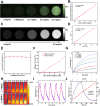
PA/MR signals and photothermal properties in vitro. (A) Concentration-dependent PA images of IMPP-c dispersions at a wavelength of 808 nm. (B) The corresponding linear regression curve illustrating the relationship between PA intensity and concentrations of IMPP-c. (C) MR images of IMPP-c dispersions at varying concentrations. (D) Relaxation rates 1/T1 (s-1) of IMPP-c as a function of Mn2+ concentrations (mM). (E) Stability test for PA signals from IMPP-c NPs under continuous 808-nm laser irradiation over a duration of 30 minutes. (F) Comparison of relative singlet oxygen (1O2) levels detected by SOSG in groups with and without laser irradiation. (G) Temperature elevation curves for IMPP-c NPs across different concentrations (0 - 0.4 mg mL−1) during exposure to laser irradiation at 808 nm with an intensity of 0.8 W·cm⁻². (H) Infrared thermal images depicting IMPP-c at a concentration of 0.4 mg mL−1 and deionized water in quartz cuvettes after being subjected to 10 minutes of 808-nm laser irradiation. (I) Photothermal heating and cooling cycles observed for IMPP-c NPs upon exposure to an 808-nm laser source. (J) Comprehensive heating and cooling profiles along with linear regression analysis for determining the time constant τs.
Figure 4
Figure 4
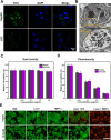
Cellular uptake and dark/photo toxicity in cell lines. (A) Confocal laser scanning microscopy images of HepG2 and LO2 cells treated with IMPP-c solutions for 4 hours. The cytoplasm exhibits green fluorescence from IMPP-c, while the cell nuclei are stained blue with DAPI, as shown in the merged image. (B) TEM images of HepG2 cells incubated with IMPP-c NPs. (C) Cell viability assessments of HepG2 and HCCLM3 cells following incubation with varying concentrations of IMPP-c NPs in darkness for 24 hours. (D) Cell viability evaluations of HepG2 and HCCLM3 cells after incubation with different concentrations of IPP-c NPs under laser irradiation for 10 minutes at a wavelength of 808 nm and an intensity of 0.8 W·cm-2. (E) Double staining images using Calcein AM (green, indicating live cells) and PI (red, indicating dead cells), depicting HepG2 and HCCLM3 cells treated with PBS, ICG, and IMPP-c both with and without laser irradiation.
Figure 5
Figure 5
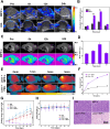
In vivo PA/MR imaging and phototherapy of subcutaneous tumors. (A) In vivo PA imaging of subcutaneous tumors at various post-injection time points following the administration of IMPP-c NPs and ICG (Excitation wavelength: 808 nm). (B) Corresponding quantification of PA signals at the tumor site in both the IMPP-c NPs and ICG injection groups. (C) In vivo MR images of mice bearing subcutaneous tumors after receiving injections of IMPP-c NPs. (D) Quantitative analysis of average MR signal intensities. (E) Infrared thermal imaging of subcutaneous HepG2-tumor-bearing mice treated with either PBS or IMPP-c, subjected to 808-nm laser irradiation (0.8 W·cm−2) for varying durations. (F) Heating curves recorded at the tumor site during laser irradiation. (G) Tumor growth curves (n = 4, mean ± SD) across different treatment groups. (H) Changes in body weight among nude mice within various treatment cohorts (n = 4). (I) Histological microscopy images depicting tumors after treatments administered across different groups over a period of 15 days. *** P<0.001.
Figure 6
Figure 6
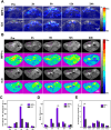
In vivo PA/MR imaging of orthotopic tumors. (A) In vivo PA imaging of the orthotopic tumor at various post-injection time points following administration of IMPP-c NPs and ICG (Excitation wavelength: 808 nm). (B) In vivo MR imaging of the orthotopic tumor at different post-injection time intervals for IMPP-c NPs and ICG. (C) Corresponding quantification of PA signals at the tumor site from (A). (D) Quantitative analysis of average MR signal intensities from (B). (E) Quantification of mean ex vivo PA signals from the liver tumor and major organs 6 hours after injection of IMPP-c NPs/ICG. *** P<0.001.
Figure 7
Figure 7
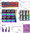
In vivo PA/MR imaging-guided phototherapy of orthotopic tumors. (A) Infrared thermal imaging was conducted on orthotopic SHCC tumor-bearing mice treated with either IMPP-c or PBS, followed by 808-nm laser irradiation (0.8 W·cm⁻²) for varying durations. (B) Heating curves of the tumor sites in both groups during laser irradiation were recorded. (C) Continuous monitoring of the MR signals from orthotopic SHCCs in both groups was performed before and after phototherapy at Pre, Day 7, and Day 14. (D) Bioluminescence signals from orthotopic SHCCs were continuously monitored in both groups before and after phototherapy at Pre, Day 7, and Day 14. (E) Relative MR signal changes of orthotopic SHCCs derived from (C). (F) Changes in body weight among mice across different treatment groups are illustrated (n = 4). (G) H&E staining results of SHCCs dissected from treated mice in both groups are presented, collected 15 days post PTT/PDT treatment. *** P<0.001.

Similar articles

References

    1. Siegel RL, Giaquinto AN, Jemal A. Cancer statistics, 2024. CA Cancer J Clin. (2024) 74:12–49. doi: 10.3322/caac.21820 - DOI - PubMed
    1. Kelley RK, Greten TF. Hepatocellular carcinoma - origins and outcomes. N Engl J Med. (2021) 385:280–2. doi: 10.1056/NEJMcibr2106594 - DOI - PubMed
    1. Llovet JM, Willoughby CE, Singal AG, Greten TF, Heikenwalder M, El-Serag HB, et al. Nonalcoholic steatohepatitis-related hepatocellular carcinoma: pathogenesis and treatment. Nat Rev Gastroenterol Hepatol. (2023) 20:487–503. doi: 10.1038/s41575-023-00754-7 - DOI - PMC - PubMed
    1. Sharpton SR, Loomba R. Emerging role of statin therapy in the prevention and management of cirrhosis, portal hypertension, and hcc. Hepatology. (2023) 78:1896–906. doi: 10.1097/HEP.0000000000000278 - DOI - PubMed
    1. Pinter M, Jain RK, Duda DG. The current landscape of immune checkpoint blockade in hepatocellular carcinoma: a review. JAMA Oncol. (2021) 7:113–23. doi: 10.1001/jamaoncol.2020.3381 - DOI - PMC - PubMed

MeSH terms

LinkOut - more resources