Review
. 2025 Dec;17(1):2519703.
doi: 10.1080/19490976.2025.2519703. Epub 2025 Jun 26.

Gastrointestinal inflammation and cancer: viral and bacterial interplay

Affiliations
Review

Gastrointestinal inflammation and cancer: viral and bacterial interplay

Valentina Arrè et al. Gut Microbes. 2025 Dec.

Abstract

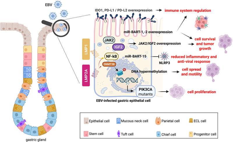
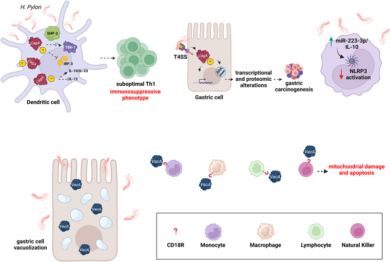
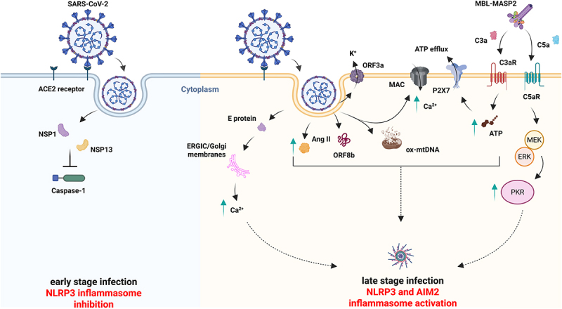
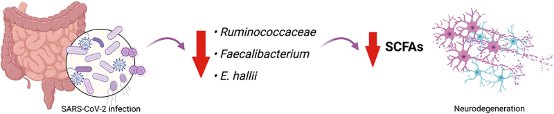
Gastrointestinal (GI) inflammation and malignancies arise from complex interactions between the host's immune responses and microbial pathogens. Epstein-Barr virus (EBV), Helicobacter pylori (H. pylori), and Severe acute respiratory syndrome coronavirus 2 (SARS-CoV-2) contribute to chronic GI inflammation, immune evasion, and tumorigenesis through distinct but interconnected mechanisms. EBV, a widespread herpesvirus, establishes a latent infection in B cells and epithelial cells. It promotes gastric carcinogenesis through immune modulation, epigenetic changes, and viral microRNAs (miRNAs). H. pylori, a gastric carcinogen, induces chronic gastritis and gastric cancer (GC) through Cytotoxin-associated gene A (CagA) and Vacuolating cytotoxin gene A (VacA) virulence factors. These factors disrupt host immune responses and enhance oncogenic signaling pathways. Recent evidence also links SARS-CoV-2 to gut dysbiosis and inflammatory responses. It worsens immune dysfunction and hence potentially impacting GI pathology. EBV and H. pylori co-infections may synergistically amplify inflammatory signaling, creating a tumor-promoting microenvironment. This review emphasizes the molecular mechanisms by which these pathogens contribute to GI diseases, focusing on their immune evasion strategies and potential therapeutic targets. Understanding these interactions is essential for developing targeted interventions for infection-driven GI malignancies.

Keywords: EBV; H. Pylori; NLRP3; SARS-CoV-2; cancer; gastrointestinal disease; immune escape; inflammation; microRNA.


Conflict of interest statement

The authors declare the absence of any commercial or financial conflict of interest.

Figures

Figure 1.
Figure 1.
EBV infection of gastric epithelial cells impinges several molecular pathways leading to GC. EBV expresses latent membrane proteins-1 and − 2A (LMP1 and LMP2A) to maintain latency and cell proliferation through a positive feedback loop with NF-kB. LMP2A activates DNA methyltransferase 3 beta (DNMT3B) causing genome-wide methylation contributing to gastric cancer (GC). The virus may promote DNA hypermethylation in GC, with phosphatidylinositol-4,5-Bisphosphate 3-kinase Catalytic Subunit alpha (PIK3CA) mutations. These mutations may lead to cell proliferation. Other pathways leading to tumor growth, induced by EBV, are Janus kinase 2 (JAK2) and insulin-like growth factor 2 (IGF2) signaling. EBV-encoded microRNA (miRNA) clusters, including miR-BART-1 and − 2, are highly expressed and promote cell survival and immune evasion. In the tumor immune microenvironment (TIME), B cells infected by EBV were found to deliver exosomes carrying miR-BART-15 to epithelial non-EBV-infected cells. The virus might utilize the same mechanism to deliver miR-BART-15 into gastric epithelial cells to inhibit the activation of the inflammasome nucleotide-binding oligomerization domain, leucine-rich repeat-containing protein 3 (NLRP3), thus mitigating the inflammatory antiviral responses. EBV evades immune recognition by overexpressing the co-inhibitors indoleamine 2,3-dioxygenase 1 (IDO1), Programmed Death-Ligand 1 and 2 (PD-L1 and PD-L2).
Figure 2.
Figure 2.
H. pylori strategies against host immune response. H. pylori infects the dendritic cells (DCs) and expresses cytotoxin-associated gene a (CagA) oncoproteins which undergo phosphorylation and activate src homology two domain-containing protein tyrosine phosphatase-2 (SHP-2). This activation suppresses TGF-beta-activated kinase 1 (TAK1) binding protein 1 (TBK-1) activation, inhibiting interferon regulatory factor-3 (IRF-3) phosphorylation and nuclear translocation. Consequently, interferon production by DCs is reduced. The bacterium proliferates within DCs, impairing their function by inhibiting the production of the pro-inflammatory cytokine IL-12. On the other hand, CagA can increase the anti-inflammatory IL-10 production, resulting in suboptimal T helper 1 (Th1) development and activation. The CagA protein is delivered into gastric epithelial cells through the bacterium’s type IV secretion system (T4SS). Once inside the host cells, CagA becomes tyrosine-phosphorylated by host kinases and interacts with various host signaling pathways, leading to altered genomic instability, inflammation, and gastric cancer. H. pylori virulence factor Vacuolating cytotoxin gene a (VacA) causes vacuolization in epithelial cells of the gastric mucosa, binds to the Cluster of differentiation 18 CD18 receptors of immune cells like monocytes, macrophages, polymorphonuclear lymphocytes (PMNs), and natural killer (NK) cells, inducing mitochondrial damage and apoptosis. H. pylori infection of a monocyte/macrophage human cell line, named THP-1, induced miR-223-3p and IL-10 expression to regulate the activation of nucleotide-binding oligomerization domain, leucine rich repeat and pyrin domain containing 3 (NLRP3) inflammasome. This mechanism fine-tunes the host inflammatory response, preventing excessive inflammation and offers novel insights into how H. pylori establishes and maintains chronic infections.
Figure 3.
Figure 3.
SARS-CoV-2 infection implies NLRP3 inflammasome attenuation at the early stage of infection while necessitating its activation to disseminate at a later stage. Angiotensin-Converting enzyme 2 (ACE2) receptors expressed on the enterocyte’s surface allow SARS-CoV-2 propagation within the gastrointestinal (GI) tract. During early-stage infection, SARS-CoV-2 employes non-structural protein 1 and 13 (NSP1 and NSP13) to deceive the host immune system by dampening the NLRP3 inflammasome activation (left panel). However, viral particles dissemination throughout late-stage infection implies the activation of the NLRP3 inflammasome, encouraging the cytokine storm and triggering systemic inflammation by employing several different strategies. The E protein sustains NLRP3 inflammasome activation through Ca2+ mobilization from intracellular stores, whereas the open reading frame 3a (ORF3a) does so via K+ efflux. Similarly, angiotensin II (Ang II) cytosolic accumulation activates the inflammasome indirectly. Moreover, active open reading frame 8b (ORF8b) physically interacts with the NLRP3 leucine-rich repeats (LRR) domain to induce its activation. Not surprisingly, late-stage infection is characterized by an active complement cascade, whose intermediates, such as membrane attack complex (MAC), complement Component 3a and 5a (C3a and C5a), converge, leading to the NLRP3 activation. MAC forms plasma membrane pores and generates Ca2+ influx. C3a ionophores enhances adenosine triphosphate (ATP) levels and promotes the ATP efflux through the Purinergic receptor P2X, ligand-gated ion channel 7 (P2X7) receptor and, then C5a signals to the mitogen-activated protein kinase (Mek)/extracellular signal-regulated kinase 1/2 (ERK 1/2)/protein kinase R (PKR) pathway, triggering NLRP3 activation. Finally, SARS-CoV-2-induced cytosolic mitochondrial DNA (mtDNA) catalyzes the NLRP3 and absent in melanoma 2 (AIM2) inflammasome activation (right panel).
Figure 4.
Figure 4.
EBV infection contributes to the onset of neurological disorders. EBV infection interferes with the host’s gut–brain axis physiology and may underpin the development of neurological disorders such as Alzheimer’s disease (AD), Parkinson’s disease (PD), and multiple sclerosis (MS). A leaking blood brain barrier (BBB) allows EBV particles to access the brain, where they instigate leukocytes and lymphocytes to produce and secrete tumor necrosis factor-α (TNF-α), which, in turn, is responsible for the hyperphosphorylation of tubulin associated unit (tau) protein, leading to neurofibrillary tangles, and the formation of amyloid β-plaques, thus disintegrating microtubules and accelerating neuron degeneration and the onset of AD (upper panel). Moreover, the viral latent membrane proteins-1 (LMP1) protein carried by infiltrating EBV can lead to dopaminergic neuron degeneration through the interaction between its DPDN sequence and α-syn, causing aggregation of the latter into protofibril structures and subsequently driving the onset of PD (middle panel). Acute EBV infection has also been linked to the onset of MS. Although the mechanisms remain unclear, molecular mimicry between EBV-encoded and mammalian-encoded proteins has recently been proposed as a potential determinant. Virally encoded Epstein–Barr virus nuclear antigen 1 (EBNA1), BamHI fragment rightward open reading frame 3 (BFRF3), and LMP1 share similarities with mammalian proteins myelin basic protein (MBP) and anoctamin 2 (ANO2), thus exposing the latter to the host antibodies and cytokines, which ultimately leads to demyelination, altered axonal electrical conduction, neuron degeneration, and MS development (lower panel).
Figure 5.
Figure 5.
Acute H. pylori infection promotes the onset of neurological disorders through inflammasome activation. Persistent H. pylori colonization might underly the onset and development of Alzheimer’s disease (AD) and Parkinson’s disease (PD). Cytotoxin-associated gene a (CagA)- signal transducer and activator of transcription 3 (STAT3)-inflammasome axis engagement results in the secretion of mature IL-1β, reaching the brain district through the vagus nerve and supporting the expression of AD-associated markers, such as Apolipoprotein E4 (ApoE4), amyloid β-precursor protein (APP), beta-site APP cleaving enzyme 1 (BACE1), and presenilin-1 (PSEN1), left panel. Moreover, besides its function in inducing inflammasome activation, LPS sustains α-syn conformational changes into protofibril structures, which, once within the neural compartment, prompt activated astrocytes and microglia to release nitric oxide (NO) and reactive oxygen species (ROS), ultimately damaging dopaminergic neurons, causes of PD onset.
Figure 6.
Figure 6.
SARS-CoV-2 infection leads to gut dysbiosis, reduction of SCFAs-producing bacteria and neurodegeneration. SARS-CoV-2-infected people show a significant decrease of SCFAs-producing bacterial species, such as Ruminococcaceae, Faecalibacterium and Eubacterium hallii (E. hallii), whose loss is responsible for neurodegeneration and brain issues.

References

    1. Burd EM, Hinrichs BH.. Gastrointestinal infections. Mol Pathol Clin Pract. 2015; 707–30. doi: 10.1007/978-3-319-19674-9_50. - DOI
    1. Quaglio AEV, Grillo TG, De Oliveira ECS, Di Stasi LC, Sassaki LY. Gut microbiota, inflammatory bowel disease, and colorectal cancer. World J Gastroenterol. 2022;28(30):4053–4060. doi: 10.3748/wjg.v28.i30.4053. - DOI - PMC - PubMed
    1. Ayoubian H, Ludwig N, Fehlmann T, Menegatti J, Gröger L, Anastasiadou E, Trivedi P, Keller A, Meese E, Grässer FA, et al. Epstein-Barr virus infection of cell lines derived from diffuse large B-Cell lymphomas alters MicroRNA loading of the Ago2 complex. J Virol. 2019. Jan 17. 93(3):e01297–18. doi: 10.1128/JVI.01297-18. - DOI - PMC - PubMed
    1. Lisaza H, Nanbo A, Nishikawa J, Jinushi M, Yoshiyama H. Epstein-Barr virus (EBV)-associated gastric carcinoma. Viruses. 2012;4(12):3420–3439. doi: 10.3390/v4123420. - DOI - PMC - PubMed
    1. Hirabayashi M, Traverse-Glehen A, Combes JD, Clifford GM, de Martel C. Estimating the prevalence of Epstein-Barr virus in primary gastric lymphoma: a systematic review and meta-analysis. Infect Agent Cancer. 2023;18(1). doi: 10.1186/s13027-023-00482-2. - DOI - PMC - PubMed

MeSH terms

LinkOut - more resources