

Midlife estradiol treatment reduces the firing rate of liver-related PVN neurons in ovariectomized high-fat diet-fed mice
- PMID: 40568886
- PMCID: PMC12288935
- DOI: 10.1152/ajpregu.00117.2025
Midlife estradiol treatment reduces the firing rate of liver-related PVN neurons in ovariectomized high-fat diet-fed mice
Abstract
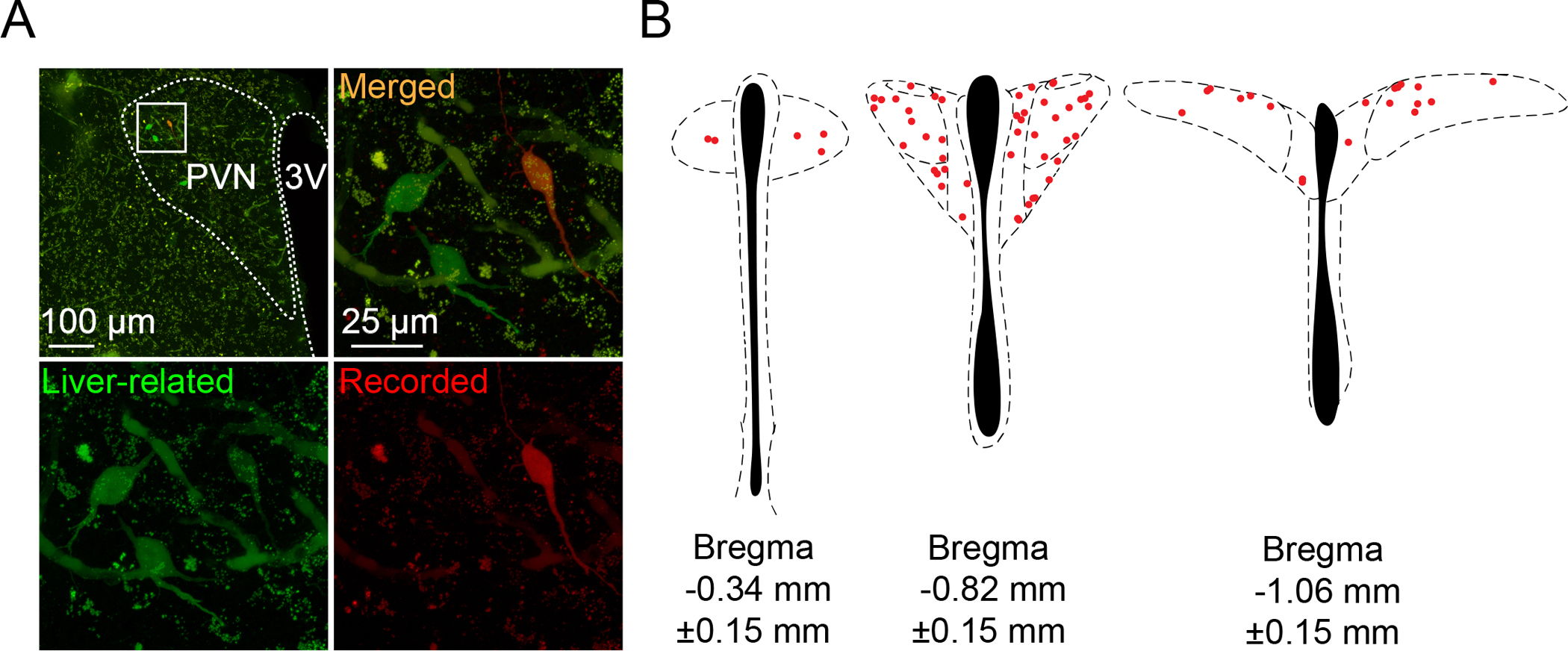
Estrogen plays a critical role in the regulation of physiological functions, including metabolism, and its involvement in the regulation of insulin sensitivity and glucose homeostasis has major clinical relevance. Despite the importance of the brain-liver pathway in the regulation of glucose metabolism and that postmenopausal women have an increased risk of developing metabolic disorders, the effect of hormone therapy on hypothalamic neurons involved in the regulation of liver metabolism is not known. Here, we tested the hypothesis that in middle-aged, high-fat diet (HFD)-fed female mice, the excitability of liver-related neurons in the paraventricular nucleus (PVN) of the hypothalamus is increased, whereas estradiol treatment attenuates this increase. Mice fed with phytoestrogen-free control (low-fat diet) or HFD were ovariectomized, received a silastic capsule implant containing either estradiol or vehicle, and stayed on their respective diets. Estradiol treatment resulted in less fat mass and lower body weight. Liver-related neurons were identified with a retrograde, transsynaptic viral tracer, and patch-clamp recordings were conducted from identified neurons in the PVN. Our data show that the excitability of liver-related PVN neurons was increased in ovariectomized HFD mice compared with LFD-fed mice. In estradiol-treated HFD mice, the firing of liver-related PVN neurons was significantly reduced compared with vehicle-treated HFD mice, whereas in LFD mice, estradiol treatment did not alter the activity of liver-related PVN neurons. Our findings suggest that midlife estradiol treatment has beneficial effects on liver-related PVN neurons and thus may contribute to the improved metabolic status observed in estradiol-treated HFD mice.NEW & NOTEWORTHY Menopause increases the risk of metabolic disorders, and despite the importance of the brain-liver pathway in the regulation of glucose homeostasis, the effect of estradiol treatment on liver-related neurons is not known. Our data show that in middle-aged, high-fat diet-fed, ovariectomized female mice, the excitability of liver-related neurons in the paraventricular nucleus is increased, whereas estradiol treatment attenuates this increase. These data suggest that midlife estradiol treatment is beneficial for the brain-liver pathway.
Keywords: electrophysiology; estradiol; high-fat diet; liver-related PVN neurons; ovariectomy.
Conflict of interest statement
Declaration of interest
None.
Figures


Similar articles
-
A subset of neurons in the paraventricular nucleus of the hypothalamus directly project to liver-related premotor neurons in the ventrolateral medulla.Auton Neurosci. 2025 Feb;257:103222. doi: 10.1016/j.autneu.2024.103222. Epub 2024 Nov 30. Auton Neurosci. 2025. PMID: 39647176 Free PMC article.
-
Selective ERα Attenuates Hypothalamic ER Stress and Regulates Energy Homeostasis in Ovariectomized Mice Fed With High-Fat Diet.J Biochem Mol Toxicol. 2025 Sep;39(9):e70446. doi: 10.1002/jbt.70446. J Biochem Mol Toxicol. 2025. PMID: 40838662
-
Enhanced fatty acid oxidation in osteoprogenitor cells provides protection from high-fat diet induced bone dysfunction.J Bone Miner Res. 2025 Feb 2;40(2):283-298. doi: 10.1093/jbmr/zjae195. J Bone Miner Res. 2025. PMID: 39657629 Free PMC article.
-
Systemic pharmacological treatments for chronic plaque psoriasis: a network meta-analysis.Cochrane Database Syst Rev. 2021 Apr 19;4(4):CD011535. doi: 10.1002/14651858.CD011535.pub4. Cochrane Database Syst Rev. 2021. Update in: Cochrane Database Syst Rev. 2022 May 23;5:CD011535. doi: 10.1002/14651858.CD011535.pub5. PMID: 33871055 Free PMC article. Updated.
-
The Black Book of Psychotropic Dosing and Monitoring.Psychopharmacol Bull. 2024 Jul 8;54(3):8-59. Psychopharmacol Bull. 2024. PMID: 38993656 Free PMC article. Review.
References
-
- Lovejoy JC, Sainsbury A, and Stock Conference Working G, Sex differences in obesity and the regulation of energy homeostasis. Obes Rev, 2009. 10(2): p. 154–67. - PubMed
MeSH terms
Substances
Grants and funding
LinkOut - more resources
Full Text Sources