. 2025 Jul 9;147(27):23781-23796.
doi: 10.1021/jacs.5c06044. Epub 2025 Jun 26.

Biosynthesis of Biphenomycin-like Macrocyclic Peptides by Formation and Cross-Linking of Ortho-Tyrosines

Affiliations

Biosynthesis of Biphenomycin-like Macrocyclic Peptides by Formation and Cross-Linking of Ortho-Tyrosines

Chandrashekhar Padhi et al. J Am Chem Soc. .

Abstract

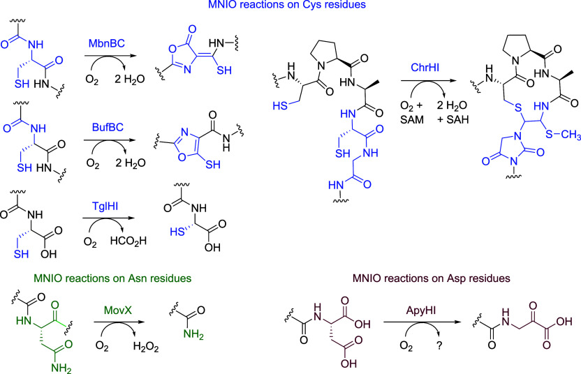
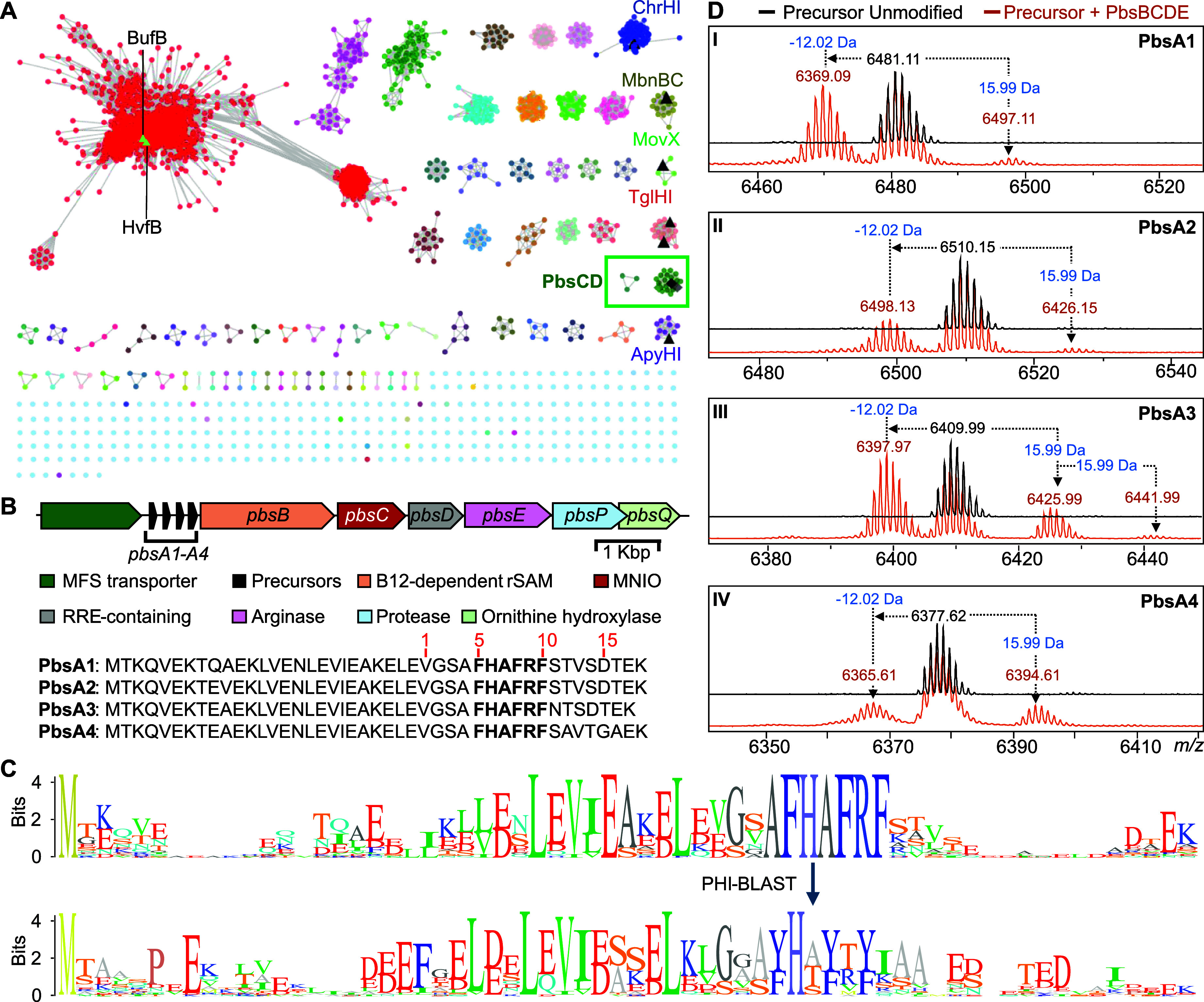
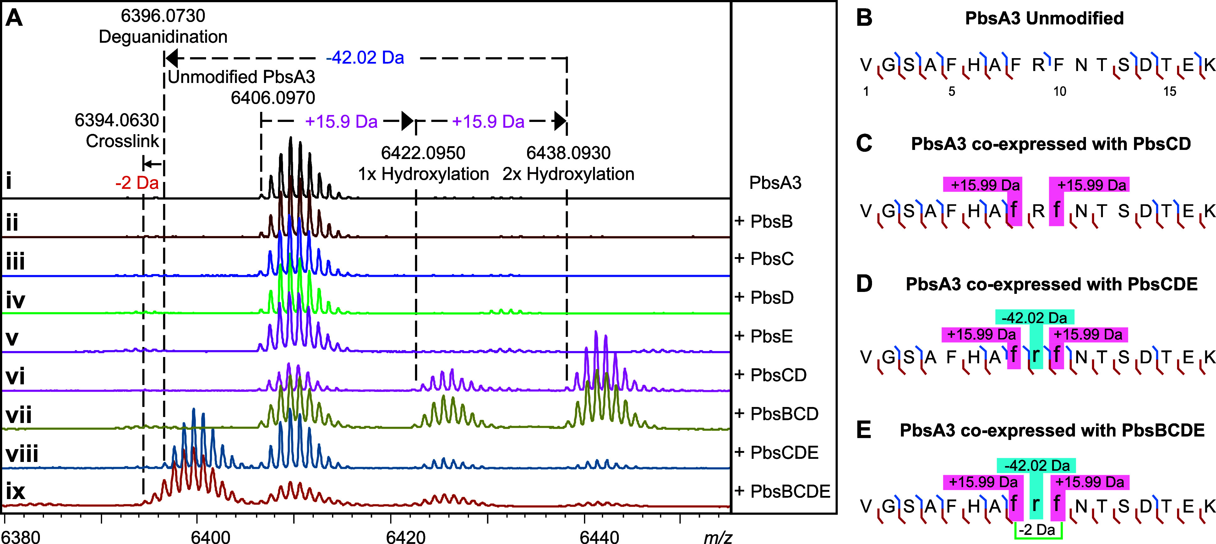
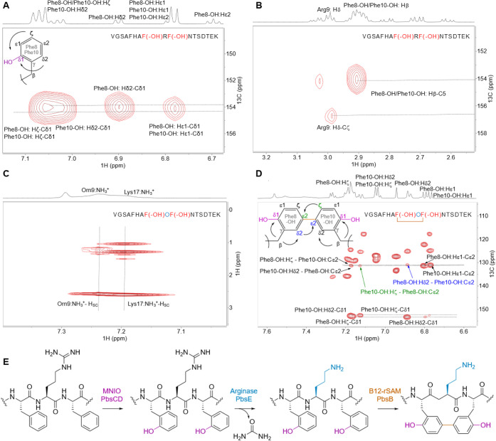
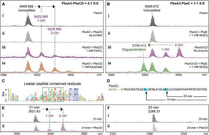
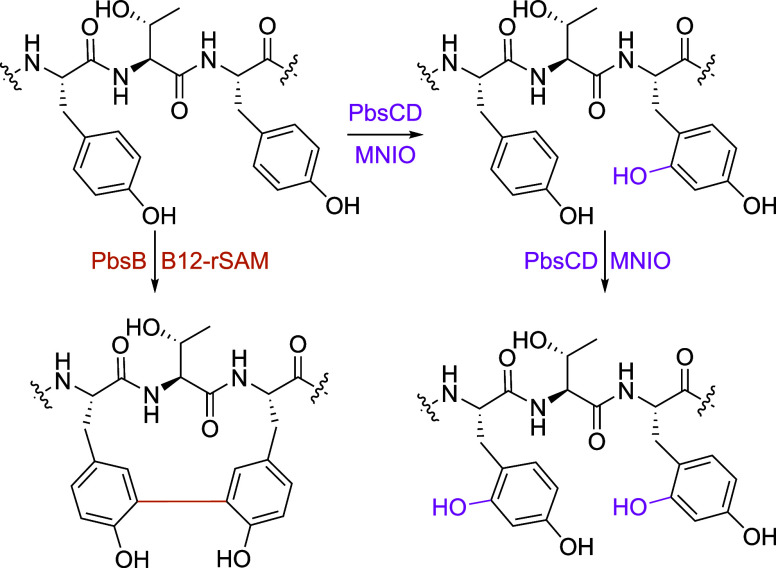
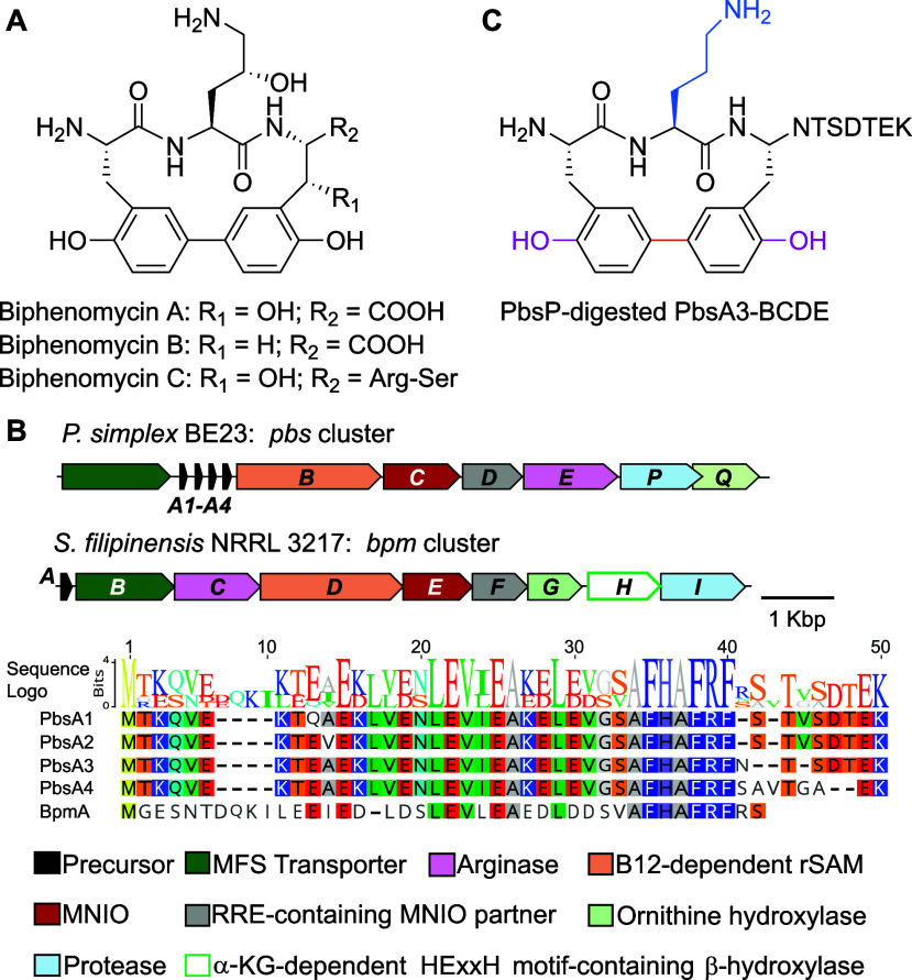
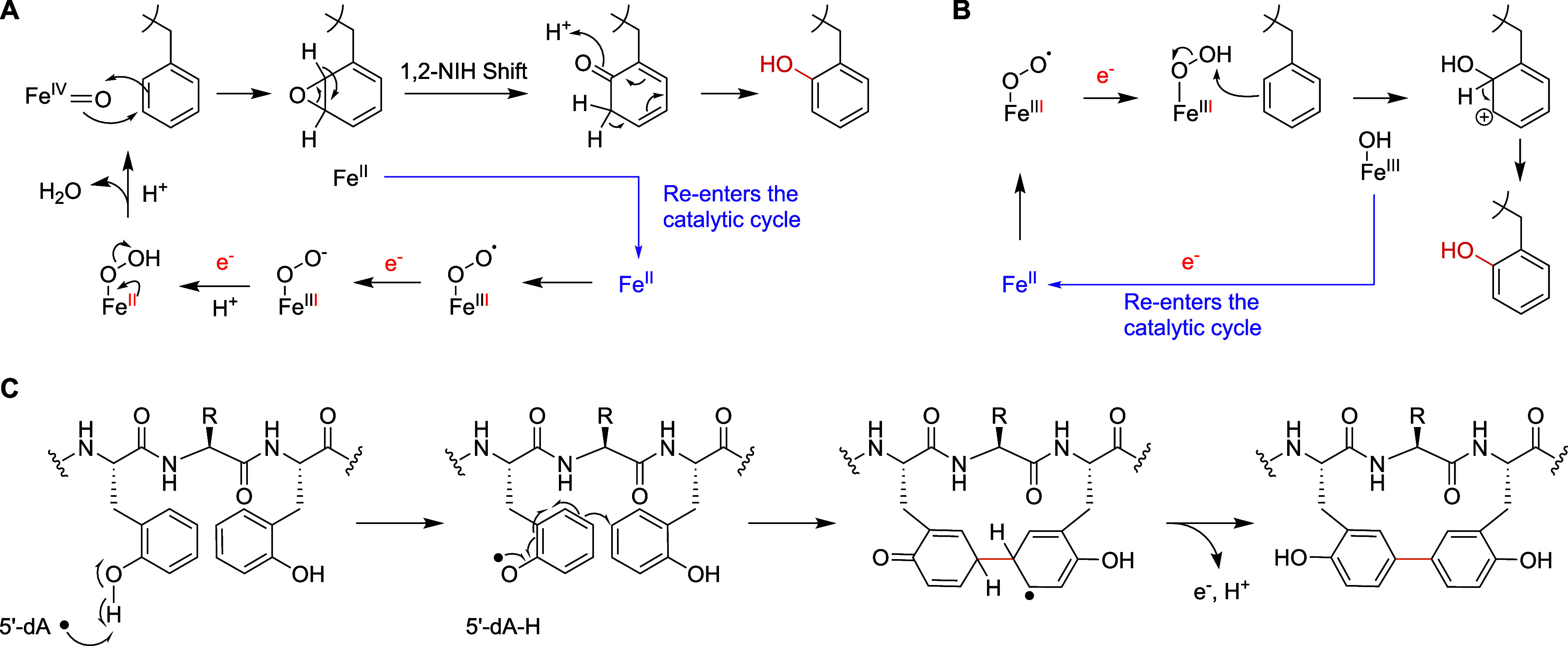
Ribosomally synthesized and posttranslationally modified peptides (RiPPs) are a growing class of natural products. Multinuclear nonheme iron-dependent oxidative enzymes (MNIOs, previously DUF692) are involved in a range of unprecedented biochemical reactions. Over 13,500 putative MNIO-encoding biosynthetic gene clusters (BGCs) have been identified by sequence similarity networks. In this study, we investigated a set of precursor peptides containing a conserved FHAFRF motif in MNIO-encoding BGCs. These BGCs contain genes encoding an MNIO, a RiPP recognition element-containing protein, an arginase, a hydroxylase, and a vitamin B12-dependent radical SAM enzyme (B12-rSAM). Using heterologous reconstitution of a representative BGC from Peribacillus simplex (pbs cluster) in E. coli, we demonstrated that the MNIO in conjunction with the partner protein catalyzes ortho-hydroxylation of each of the phenylalanine residues in the conserved FRF motif, the arginase forms an ornithine from the arginine, the ornithine residue is hydroxylated, and the B12-rSAM cross-links the ortho-Tyr side chains by a C-C linkage forming a macrocycle. A protease matures the RiPP to its final form. The elucidated structure shares close similarity to biphenomycins, a class of peptide antibiotics for which the biosynthetic pathway has not been characterized. Substrate scope studies suggest some tolerance of the MNIO and the B12-rSAM enzymes. This study expands the diverse array of posttranslational modifications catalyzed by MNIOs and B12-rSAM enzymes, deorphanizes biphenomycin biosynthesis, and provides a platform for the production of analogs from orthologous BGCs.


Figures

1
1
MNIO catalyzed reactions. The majority of known MNIO reactions utilize Cys residues as the substrate (in blue). Only two noncysteine-based reactions acting on Asn (green) and Asp (maroon) residues have been reported to date.
2
2
Genome mining of MNIO-containing BGCs encoding precursors with conserved motifs. (A) An SSN of ca. 13,500 MNIO homologues was generated with the EFI tools using the UNIREF90 database. RepNode 50 is shown as visualized in cytoscape v3.10. Previously characterized MNIOs are depicted in black triangles. The lime-colored triangles represent the nodes for HvfB and BufB that remain underneath the other nodes in the red cluster. The cluster boxed in green contains the MNIO PbsC that is the focus of this study. (B) The pbs gene cluster with the amino acid sequences of the precursors PbsA1-A4. The numbering in red is based on the GluC-digested peptide fragment isolated for further structural characterization (vide infra). (C) Sequence logo of precursor peptides containing the conserved FHAFRF motif, extracted from orthologous BGCs in the SSN (box in panel A). (D) MALDI-TOF mass spectra of N-terminally 6xHis-tagged PbsA1-A4 peptides expressed alone (black trace) or coexpressed with PbsB, PbsC, PbsD, and PbsE in (orange trace); subpanels assigned to Roman numerals I–IV for PbsA1–A4, respectively.
3
3
Mass spectrometric analysis of PbsA3 coexpressed with PbsB, PbsC, PbsD, and PbsE in different combinations. (A) MALDI-TOF MS spectra of the PbsA3 peptide when expressed (i) alone or coexpressed with (ii) the B12-rSAM, PbsB; (iii) the MNIO, PbsC; (iv) the RRE-containing partner protein, PbsD; (v) the arginase, PbsE; (vi) PbsC and PbsD; (vii) PbsB, PbsC, and PbsD; (viii) PbsC, PbsD, and PbsE; and (ix) PbsB, PbsC, PbsD, and PbsE. HR-MS/MS fragmentation patterns for the endopeptidase GluC-digested peptide fragments are shown for (B) unmodified PbsA3, (C) PbsA3 coexpressed with PbsCD, (D) PbsA3 coexpressed with PbsCDE, and (E) PbsA3 coexpressed with PbsBCDE. The peptide fragments after endoproteinase GluC digestion are numbered starting at Val1 (Figure B) with b-ions displayed in blue and y-ions displayed in red. The residues undergoing mass changes are shown in small font, further highlighted in boxes with their respective mass shifts. The hypothesized cross-linking residues are joined by a green bracket in panel E. For ESI MS/MS spectra, see Figure S4.
4
4
Structure determination and biosynthetic pathway of modified PbsA3 digested with endoproteinase GluC. (A) 1H–13C HMBC spectrum of PbsA3-CD highlighting the ortho-hydroxylated Phe8-OH and Phe10-OH residues. (B) 1H–13C HMBC spectrum highlighting the cross-peaks between the β-protons of Phe8-OH and Phe10-OH and their respective hydroxylated aromatic carbons. This part of the spectrum also shows a diagnostic cross peak for Arg9 that is consistent with an unmodified residue. (C) 1H–1H TOCSY spectrum of PbsA3-CDE collected at 3 °C in 0.1% formic acid highlighting the cross peaks of the NH3 + moiety on the Cδ of the residue at position 9 with the other side chain protons (labeled as Hsc). (D) 1H–13C HMBC correlations in PbsA3-BCDE highlighting the cross-link formed between Cε2 carbons of the ortho-hydroxylated Phe8-OH and Phe10-OH residues. Cross peaks for the Hζ of Phe10-OH to the Cε2 carbon of Phe8-OH (in green arrow/font) and for the Hδ2-proton of Phe8-OH to the Cε2 carbon of Phe10-OH (in blue arrow/font) are shown. For panels A–D, the sequences of the analyzed peptides are shown in the spectra. Modified residues are in red (hydroxylated Phe8/10) or blue font (ornithine). (E) Biosynthetic pathway of PbsA3 modified by PbsCD, followed by PbsE and PbsB.
5
5
In vitro reconstitution of MNIO and arginase activity and minimal substrate determination for the MNIO. (A) MALDI-TOF MS spectra of PbsA3 after in vitro reaction with (i) no enzyme, (ii) PbsCD, as isolated; (iii) PbsCD, in the presence of FeSO4; and (iv) PbsCD, in the presence of sodium ascorbate. (B) MALDI-TOF mass spectra of PbsA3 after in vitro reaction with (i) no enzyme and (ii) PbsE in the presence of MnSO4. No arginase activity was detected on unmodified PbsA3. MALDI-TOF mass spectra of (iii) PbsCD-modified PbsA3, containing a mixture of unmodified, singly hydroxylated and bis-hydroxylated product, and (iv) PbsA3-CD when reacted in vitro with PbsE in the presence of MnSO4. (C) Sequence logo of the FHAFRF motif containing precursors showing conserved residues in the leader peptide, highlighted in the box. (D) Partial LysC proteolysis of PbsA3 generated 20-mer and 31-mer fragments that were purified by HPLC. The 31-mer fragment contains the conserved residues of the leader peptide, which are absent in the 20-mer fragment. (E) MALDI-TOF mass spectra of the 31-mer fragment after in vitro reaction with (i) no enzyme and (ii) PbsCD in the presence of sodium ascorbate. (F) MALDI-TOF mass spectra of the 20-mer fragment after in vitro reaction with (i) no enzyme and (ii) PbsCD in the presence of sodium ascorbate.
6
6
PbsA3-YTY variant modification catalyzed by PbsBCD. The B12-rSAM enzyme PbsB-mediated cross-linking of Tyr residues in the PbsA3-YTY variant. PbsCD first hydroxylates the Tyr10 residue and then Tyr8.
7
7
Biphenomycin structure and its BGC. (A) Structures of biphenomycins A, B, and C. (B) BGC identified in one of the biphenomycin producer strains, NRRL 3217, encoding enzymes that are homologous to the Pbs enzymes characterized in this study. The corresponding precursors are aligned. (C) Structure of PbsP-digested PbsA3-BCDE displaying structural similarity with the biphenomycins.
8
8
Possible reaction mechanisms for the MNIO and B12-dependent rSAM enzyme from the pbs BGC. (A) Possible mechanism of hydroxylation catalyzed by the MNIO PbsC using an Fe­(IV)-oxo through an epoxide intermediate followed by a hydride shift (1,2-NIH shift). Other mechanisms that do not involve a hydride migration (electrophilic aromatic substitution like) can also be drawn. (B) An alternate mechanism potentially initiated by a Fe­(III)-peroxo species. (C) Proposed mechanism of C–C cross-linking between the hydroxylated Phe8 and Phe10 residues initiated by hydrogen abstraction by a 5′-deoxyadenosyl radical.

Update of

Similar articles

References

    1. Fryszkowska A., Devine P. N.. Biocatalysis in drug discovery and development. Curr. Opin. Chem. Biol. 2020;55:151–160. doi: 10.1016/j.cbpa.2020.01.012. - DOI - PubMed
    1. Wu S., Snajdrova R., Moore J. C., Baldenius K., Bornscheuer U. T.. Biocatalysis: enzymatic synthesis for industrial applications. Angew. Chem., Int. Ed. 2021;60:88–119. doi: 10.1002/anie.202006648. - DOI - PMC - PubMed
    1. Intasian P., Prakinee K., Phintha A., Trisrivirat D., Weeranoppanant N., Wongnate T., Chaiyen P.. Enzymes, in vivo biocatalysis, and metabolic engineering for enabling a circular economy and sustainability. Chem. Rev. 2021;121:10367–10451. doi: 10.1021/acs.chemrev.1c00121. - DOI - PubMed
    1. Buller R., Lutz S., Kazlauskas R. J., Snajdrova R., Moore J. C., Bornscheuer U. T.. From nature to industry: Harnessing enzymes for biocatalysis. Science. 2023;382(6673):eadh8615. doi: 10.1126/science.adh8615. - DOI - PubMed
    1. Savile C. K., Janey J. M., Mundorff E. C., Moore J. C., Tam S., Jarvis W. R., Colbeck J. C., Krebber A., Fleitz F. J., Brands J., Devine P. N., Huisman G. W., Hughes G. J.. Biocatalytic asymmetric synthesis of chiral amines from ketones applied to sitagliptin manufacture. Science. 2010;329:305–309. doi: 10.1126/science.1188934. - DOI - PubMed

MeSH terms

LinkOut - more resources