. 2025 Jun 20;53(12):gkaf550.
doi: 10.1093/nar/gkaf550.

Unique conformational dynamics and protein recognition of A-to-I hyper-edited dsRNA

Affiliations

Unique conformational dynamics and protein recognition of A-to-I hyper-edited dsRNA

Christoph Müller-Hermes et al. Nucleic Acids Res. .

Abstract

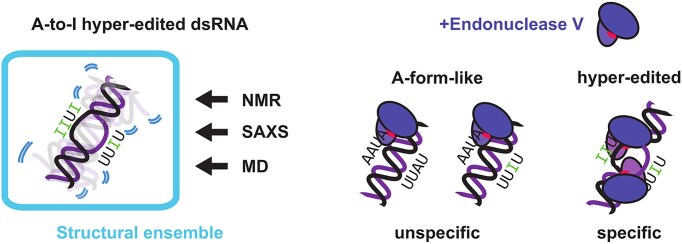
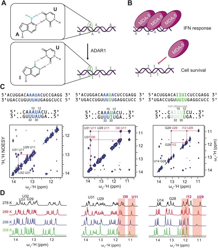
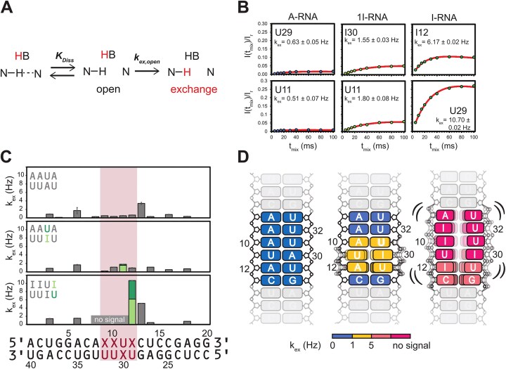
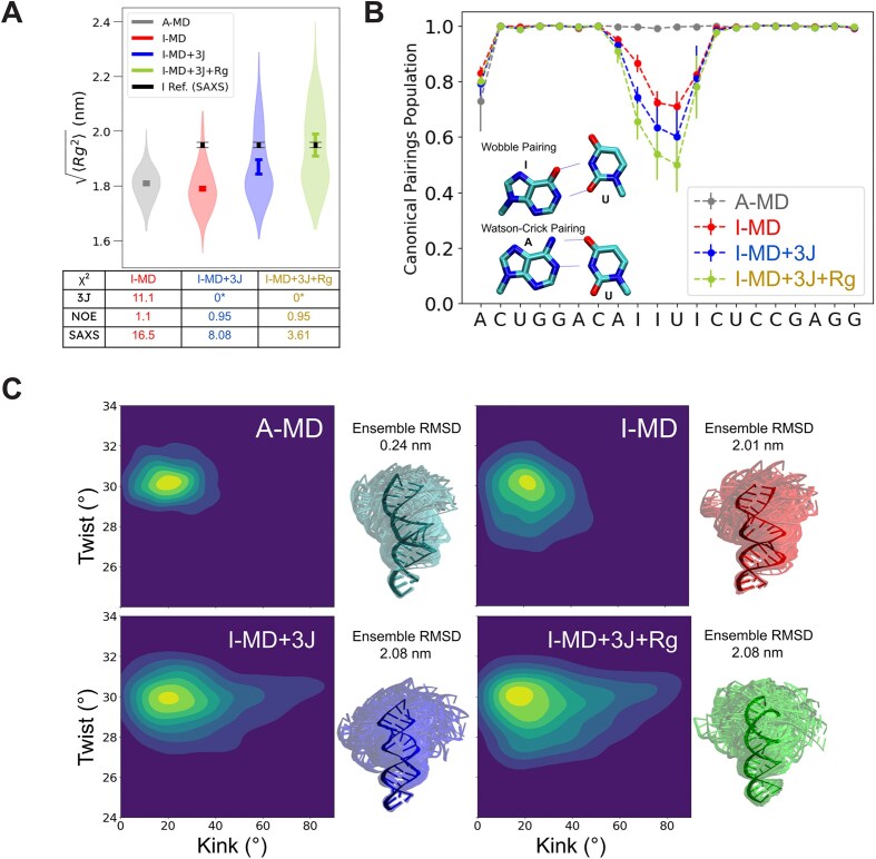
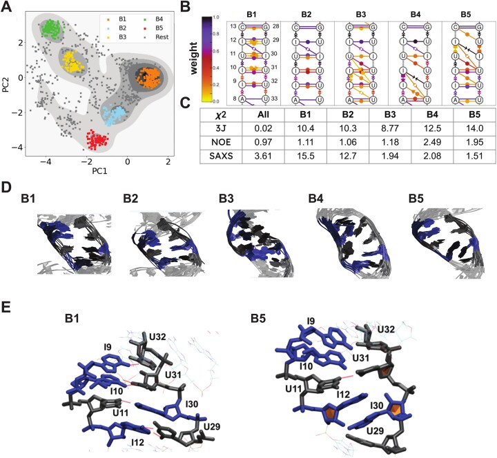
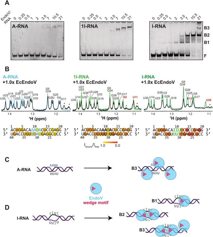
Adenosine-to-inosine (A-to-I) editing is a highly abundant modification of double-stranded RNA (dsRNA) and plays an important role in posttranscriptional gene regulation. Editing of multiple inosines by the ADAR1 enzyme leads to A-to-I hyper-editing of non-coding dsRNA, such as 3'UTRs, transposable elements, or foreign pathogenic RNAs, and is implicated in immune response and human diseases including cancer. The structural consequences of hyper-editing and its role in protein binding are poorly understood. Here, we combine solution nuclear magnetic resonance spectroscopy (NMR), biophysical methods such as small-angle X-ray scattering, and molecular dynamics simulations to study the sequence-dependent effects on conformation and dynamics of A-to-I hyper-editing for a 20-mer dsRNA and recognition of such RNAs by Endonuclease V. By comparing non-edited, single-edited, and hyper-edited dsRNA, we identify unique conformational features and extensive dynamics associated with hyper-editing, resulting in significantly increased base-pair opening. Hyper-edited dsRNA is more extended and adopts a highly dynamic ensemble of canonical and non-canonical conformations, which lead to preferential binding by Endonuclease V. Our integrated experimental and computational analysis identifies unique structural and dynamic features that are likely linked to specific protein recognition and the unique biological consequences of hyper-editing.


Conflict of interest statement

None declared.

Figures

Graphical Abstract
Graphical Abstract
Figure 1.
Figure 1.
(A) Incorporation of inosine into dsRNA by ADAR1 leads to formation of I:U base pairs. (B) Cytosolic A-form RNA is recognized by MDA-5, which leads to an innate immune response, damaging the cell. Hyper-edited dsRNA cannot be recognized by MDA-5, preventing the immune response. (C) Model dsRNAs. 1H, 1H NOESY of the duplexes were recorded at 278 K, using a mixing time of 150 ms. For non-edited dsRNA (A-RNA) and dsRNA with a single I:U base pair (1I-RNA), a full “imino walk” was possible, indicating helicity. For hyper-edited dsRNA (I-RNA), this walk was not possible, and only the imino resonances of I12:U29 are observed. The stretch of A8-U11/I30-U33 does not show imino signals. (D) Upon increasing temperature I:U base pair imino resonances show line broadening, which is more pronounced in hyper-edited dsRNA.
Figure 2.
Figure 2.
(A) Mechanism of solvent exchange between a base catalyst (i.e. H2O or a buffer component) and an imino proton in a base pair. The base pair shows an equilibrium between open and closed states. Exchange happens from the open state. (B) CLEANEX-PM build-up curves of imino signals in the central motif of A-, 1I-, and I-RNA. Exchange parameters kex are indicated. (C) Column plot of the kex values of all signals in the three dsRNAs. The central region is indicated by a red box. (D) Cartoon representation of base-pair opening in the three dsRNAs. The values of kex are color-coded. Signals not observed in the 1D spectra are shown in dark red, indicating predominantly open states.
Figure 3.
Figure 3.
(A) C3′ endo and C2′ endo conformations lead to different values of 3J(H1′,H2′). (B) 3J(H1′,H2′) values for the residues of the central motif in I-RNA. (C13C R2 rates and {1H}-13C hetNOE values for the aromatic C8/C6 in the central motif of I-RNA. (D) NMR-derived structural models of A-RNA (blue) and I-RNA (green). (E) Pairwise distance distributions of A-RNA (blue) and I-RNA (green); Rg and Dmax values are indicated. (F) SAXS profiles of A-RNA and I-RNA, including fits and residuals for structural models.
Figure 4.
Figure 4.
(A) Distribution of radius of gyration values of different MD-generated ensembles. A-RNA (gray), I-RNA (red) (both using a RECT sampling scheme), I-RNA with RECT/ME sampling scheme using 3J(H1′,H2′) values (blue), and I-RNA with RECT/ME sampling scheme using 3J(H1′,H2′) values and Rg2 (green). The χ2 values in respect to the 3J couplings, NOEs and full SAXS spectra are listed. (B) Populations of canonical pairings of the different ensembles shown in panel (A). (C) Plots of twist angles against kink angles of the different ensembles shown in panel (A) and bouquet representations with respective RMSD values. The bouquets were generated with 100 structures randomly extracted from each ensemble and aligned with respect to the eight residues at the bottom of the helix. The centroids of the ensembles are opaque colored. The ensemble RMSDs are computed on the whole ensembles with respect to the same alignment of the bouquets, using the centroids as a reference.
Figure 5.
Figure 5.
(A) 2D density plot of a principal components analysis using torsional angles and G vectors as input. A centroid-based clustering analysis was performed. (B) Dynamic secondary structures of the five clusters shown in panel (B) using the Leontis–Westhof notation, the population of the interactions is color-coded accordingly. (C) The χ2 values for the agreement of the individual clusters for 3J, NOEs, and full SAXS spectra are shown for the individual clusters (B1–B5) and the combination of the clusters (“All”). (D) Individual structures of the five clusters (B1–B5) are overlaid in respect to the central motif. Inosines are shown in blue, and uracils are shown in black. The flanking regions are shown in light gray. (E) Conformational representative for clusters B1 and B5. A non-canonical stacking interaction between residues I12 and I30 is observed in B5.
Figure 6.
Figure 6.
(A) Electromobility binding shift assays of E. coli Endonuclease V (EndoV) with A-RNA, 1I-RNA, and I-RNA. 0.2 mM RNA was used in each lane. The free and bound RNA forms were separated on 12% native polyacrylamide gels. The free RNA is labeled with "F", bands of protein/RNA complexes are "B1", "B2", and "B3". The gels were stained using SYBR Gold. (B) Effect of EndoV binding on the imino resonances of the dsRNAs. Samples containing 50 μM of A-, 1I-, or I-RNA in 25 mM sodium phosphate pH 6.5, 150 mM NaCl, 5 mM MgCl2, 1 mM DTT, and 5% D2O were measured at 950 MHz, 288 K as a reference. EcEndoV was added in a molar ratio of 1:1. Asterisks mark protein side chain signals. The observed line broadening was plotted on the sequences. (C) Binding of EndoV to A-RNA occurs as non-specific electrostatic interactions. (D) Binding of EndoV to I-RNA has different binding modes, with two specific binding modes, B1 and B2, where binding occurs to the hyper-edited central motif as well as an unspecific binding mode, B3, which is similar to A-RNA and 1I-RNA.

Similar articles

References

    1. Eisenberg E, Levanon EY A-to-I RNA editing—immune protector and transcriptome diversifier. Nat Rev Genet. 2018; 19:473–90. 10.1038/s41576-018-0006-1. - DOI - PubMed
    1. Zhang P, Zhu Y, Guo Q et al. On the origin and evolution of RNA editing in metazoans. Cell Rep. 2023; 42:112112. 10.1016/j.celrep.2023.112112. - DOI - PMC - PubMed
    1. Hoopengardner B, Bhalla T, Staber C et al. Nervous system targets of RNA editing identified by comparative genomics. Science. 2003; 301:832–6. 10.1126/science.1086763. - DOI - PubMed
    1. Levanon EY, Eisenberg E, Yelin R et al. Systematic identification of abundant A-to-I editing sites in the human transcriptome. Nat Biotechnol. 2004; 22:1001–5. 10.1038/nbt996. - DOI - PubMed
    1. Wu B, Peisley A, Richards C et al. Structural basis for dsRNA recognition, filament formation, and antiviral signal activation by MDA5. Cell. 2013; 152:276–89. 10.1016/j.cell.2012.11.048. - DOI - PubMed

MeSH terms

LinkOut - more resources